
- About the Journal
- Editorial Board
- Review Process
- Author Guidelines
- Article Processing Charges
- Special Issues
- Current Issue
- Past Issue
 Journal of Basic and Applied Pharmaceutical Science
 Volume 3 (2025), Article ID: JBAPS-109
https://doi.org/10.33790/jbaps1100109Research Article
Potential Inhibitors for Nipah Virus Glycoprotein Identified by Virtual Screening
Pratik Khanal1*, and Bhawana Sen1
Department of Pharmacy, Karnali College of Health Science, Purbanchal University, Gaushala, Kathmandu, Nepal.
Corresponding Author Details: Pratik Khanal, Department of Pharmacy, Karnali College of Health Science, Purbanchal University, Gaushala, Kathmandu, Nepal.
Received date: 21st October, 2024
Accepted date: 12th April, 2025
Published date: 15th April, 2025
Citation: Khanal, P., & Sen, B., (2025). Potential Inhibitors for Nipah Virus Glycoprotein Identified by Virtual Screening. J Basic Appl Pharm Sci, 3(1): 109.
Copyright: ©2025, This is an open-access article distributed under the terms of the Creative Commons Attribution License 4.0, which permits unrestricted use, distribution, and reproduction in any medium, provided the original author and source are credited.
Abstract
Nipah virus (NiV) is a highly pathogenic, zoonotic virus responsible for severe respiratory and neurological diseases, with a case fatality rate between 40% and 70%. Despite its global health threat, no effective antiviral treatments have been developed to date. The glycoprotein (NiV-G), responsible for viral attachment to host cells, presents an attractive target for therapeutic intervention. In this study, we employed virtual screening techniques to identify potential inhibitors of the NiV-G protein. A comprehensive library of 3,000 small molecules was screened using Auto Dock to predict their binding affinity with the viral glycoprotein.
Top-ranked ligands were subjected to molecular dynamics (MD) simulations to assess the stability of their binding interactions over time. Among the screened compounds, three candidates—Ligand201, Ligand2701, and Ligand1883—demonstrated high binding affinities, with binding free energy in the range (-7.8 kcal/mol, -6.7 kcal/mol, and -6.7 kcal/mol, respectively). Detailed analysis of their binding modes revealed strong interactions with critical residues in the receptor-binding domain of NiV-G, potentially blocking its interaction with host cell receptors ephrin-B2 and ephrin-B3.
The identified inhibitors provide promising lead compounds for further in vitro and in vivo testing, and they represent a significant step toward the development of antiviral therapies for Nipah virus infections. Future work will focus on optimizing these candidates for drug-likeness and bioavailability.
Keywords: Nipah Virus; Niv-G; Virtual Screening; Molecular Docking, In Vitro Experiments
Introduction
Nipah virus (NiV), an emerging zoonotic pathogen classified within the Paramyxoviridae family, represents a severe and escalating threat to global public health [1]. The virus was first identified during an outbreak in Malaysia in 1999, where it was associated with severe respiratory illness and encephalitis in humans, primarily affecting those in close contact with infected pigs [2]. Since then, Nipah virus has been linked to several outbreaks across Asia, including Bangladesh and India, with significant morbidity and mortality rates [3]. The World Health Organization (WHO) has highlighted Nipah virus as a high-priority pathogen due to its potential to cause widespread epidemics and its lack of effective treatments [4].
The clinical manifestations of Nipah virus infection range from flu-like symptoms to severe neurological complications, including encephalitis and coma [5]. The case fatality rate varies from 40% to 75%, making it one of the deadliest infectious diseases known. Transmission of Nipah virus occurs primarily through contact with infected animals, particularly fruit bats, which are the natural reservoir of the virus [6]. Additionally, the virus can spread through contaminated food, particularly date palm sap, and through person to-person contact, particularly in healthcare settings and among family members of infected individuals [7].
The recent resurgence of Nipah virus outbreaks underscores the urgent need for effective therapeutic interventions. In July 2024, the Kathmandu Post reported that the Epidemiology and Disease Control Division of Nepal has issued a high alert due to the risk of a Nipah virus outbreak following recent cases in India [8]. Recent reports have highlighted Kerala, India, as one of the regions most at risk globally for Nipah virus outbreaks, with health authorities issuing alerts following the death of a 14-year-old boy, marking yet another tragic incident linked to the zoonotic virus, which has a high mortality rate and no available treatment or vaccine [9].
The glycoprotein (NiV-G) of the Nipah virus plays a critical role in the life cycle of virus. It is responsible for mediating the attachment of the virus to host cell receptors, initiating the infection process. Given its central role in viral entry, NiV-G represents a compelling target for antiviral drug development [10]. Despite its importance, there are currently no licensed vaccines or specific antiviral treatments for Nipah virus infection. The lack of effective therapeutic options underscores the need for innovative approaches to drug discovery [11].
In this context, the present study aims to identify potential inhibitors of NiV-G through computational methods, including virtual screening, molecular docking, and molecular dynamics simulations. Similar in silico approaches have successfully identified phytochemicals with potential inhibitory effects against NiV-G [12]. By exploring a diverse library of small molecules, this research seeks to uncover compounds that can effectively interact with and inhibit NiV-G. The use of virtual screening allows for the efficient evaluation of a large number of potential inhibitors, while molecular docking provides insights into their binding interactions with the glycoprotein [13]. Molecular dynamics simulations will further assess the stability and dynamics of these interactions, providing a comprehensive understanding of their potential efficacy [14].
The outcomes of this research could significantly advance the development of antiviral therapies for Nipah virus, addressing a critical gap in the current treatment landscape. Identifying and optimizing promising drug candidates will not only contribute to global health efforts in managing Nipah virus outbreaks but also provide a foundation for future research aimed at combating other emerging viral threats.
By focusing on NiV-G and leveraging advanced computational techniques, this study aims to contribute valuable insights to the field of antiviral drug discovery and support public health initiatives aimed at mitigating the impact of Nipah virus infections.
Methodology
Protein Structure Preparation
The Nipah virus glycoprotein (NiV-G), a key target in antiviral drug discovery, mediates viral entry into host cells by binding to ephrin-B2 and ephrin-B3 receptors [15]. For this study, the 3D structure of the prefusion-stabilized NiV-G (PDB ID: 3d11) was obtained from the Protein Data Bank (PDB)[15].
Protein Preprocessing Using Chimera
The protein structure was prepared for docking and molecular dynamics simulations using UCSF Chimera.
• Hydrogen Atom Addition: Hydrogens were added to the protein structure using Chimera's "Add Hydrogens" feature. The receptor preparation settings were configured as follows: hydrogens were added, charges were merged with non-polar hydrogens, and lone pairs were removed. Water molecules and chains of non-standard residues were ignored, while non-standard residues that are critical for the protein-ligand interaction were retained.
• Protonation States: The protonation states of critical residues like histidine were carefully adjusted using Chimera to account for physiological pH. Residues such as histidine, aspartic acid, and glutamic acid were assigned their most likely charged states at neutral pH. For histidine, multiple protonation states (delta, epsilon, and both forms) were considered using a residue-name based approach.
• Charge Assignment: Using the AutoDock options within Chimera, partial charges were assigned to the protein. The AMBER ff99SB force field was selected for standard residues, while the AM1-BCC charge model was applied to nonstandard residues, as shown in the second attached image. This ensured accurate electrostatic modeling for subsequent docking studies.
• Ligand Preparation: For ligand preprocessing, the following options were selected: merging charges and removing non polar hydrogens was set to true, as well as merging charges and removing lone pairs.
• Dock Prep Options: During preparation, solvent molecules were deleted, and for alternate residue locations, only the highest occupancy was retained. Specifically, the following modifications were applied: selenomethionine was changed to methionine, bromo-UMP was changed to UMP, methylselenyl dUMP was changed to UMP, and methylselenyl-dCMP was changed to CMP. Hydrogens and charges were added as part of the preparation process.
• Receptor Volume and Size: The receptor volume was set at coordinates (x: 28.21, y: 6.84, z: 90.67), with a receptor size of (x: 25.22, y: 38.94, z: 36.81), ensuring that the docking grid encompassed the binding site accurately
Energy Minimization
To resolve steric clashes and optimize the protein’s geometry, the protein structure underwent energy minimization using Chimera. The AMBER ff99SB force field was applied to standard residues to perform this step. The energy minimization converged efficiently, indicating that the protein structure was optimized and in a suitable state for molecular docking and dynamics simulations.
Ligand Library Preparation and Virtual Screening
A virtual screening approach was employed to identify potential inhibitors of NiV-G from a compound library of 3,000 small molecules, obtained from the ZINC database.
Library Preparation
The ligand library was processed using OpenBabel to ensure the 3D structures of all small molecules were properly optimized:
• Ionization and Tautomeric Forms: Ligands were processed to generate all possible ionization and tautomeric states at physiological pH (7.0). This ensures that the correct protonation states of acidic and basic groups (e.g., carboxylates, amines) are available for docking, including deprotonation of carboxylic acid (COOH to COO⁻).
• Energy Minimization: Each ligand was energy minimized to eliminate any steric clashes and ensure optimal geometries.
• Partial Charges: Gasteiger charges were added to each ligand to ensure proper charge representation for docking.
Molecular Docking
In the molecular docking process, the active site of the Nipah virus glycoprotein (NiV-G) was carefully selected based on key residues that were previously reported. These residues—Asp219, Pro220, Pro276, Asn277, Val279, Tyr280, His281, Cys282, Tyr351, Gly352, Pro353, Pro448, Phe458, Gly489, Gln490, Gly506, Val507, Tyr508, Lys560, and Gly559—were identified as crucial for interactions with ligands [16]. The grid generation for docking simulations was centered around specific coordinates, with the X-axis at 28.1 Å, Y-axis at 6.84 Å, and Z-axis at 90.67 Å. This ensured that the docking experiments targeted the precise location of the active site.
The grid box dimensions were set to cover a wide area of the protein, allowing sufficient space for the ligand binding simulations, with size parameters set as 21.44 Å in the X-axis, 33.10 Å in the Y-axis, and 31.29 Å in the Z-axis. These dimensions were chosen to include all potential interaction points within the active site. AutoDock Vina software was used for the docking protocol with an exhaustiveness level of 8, ensuring thorough sampling of the conformational space, and the number of modes (binding poses) was set to 9 [17]. Emerging transformer-based models like Dockformer have shown promise in enhancing docking accuracy and efficiency [18]. Each ligand was docked individually, and the binding affinities were used to score and rank the ligands for further analysis, ensuring the identification of potential inhibitors for the NiV-G protein.
Scoring and Ranking
AutoDock Vina evaluates the docking results based on the predicted binding affinities, represented by the binding energies in kcal/mol. These values indicate the strength of interactions between the ligand and the receptor protein. The scoring function used by Vina considers several types of molecular interactions, including hydrogen bonding, van der Waals forces, hydrophobic interactions, and electrostatic interactions. Ligands are ranked according to their binding energies, with more negative values suggesting stronger binding affinities.
Following the docking simulations, each ligand was individually docked against the NiV-G receptor, and the resulting binding energies were used to rank the ligands. The top-scoring ligands, indicating the strongest binding affinities, were selected for further analysis to identify potential inhibitors of the Nipah virus glycoprotein.
Pharmacokinetic and Toxicity Profiling
Pharmacokinetic Profiling: - All the docked molecules were evaluated for their pharmacokinetic properties:
• Lipinski's Rule of Five was applied to assess drug-likeness, focusing on molecular weight, hydrogen bond donors/acceptors and logP. The RDKit library in Python calculated these values [19].
• Veber’s Rule was used to evaluate oral bioavailability based on the number of rotatable bonds and polar surface area (PSA). The RDkit library in Python also calculated these values [20].
Toxicity Profiling: - ProTox 3.0 a virtual lab was used for the prediction of toxicities of compounds with highest binding affinity [21].
Final Selection of Hits
The top compounds were selected based on their overall docking scores, and favorable pharmacokinetic profiles. Three lead candidates— Ligand201, Ligand2701, and Ligand1883— demonstrated high binding affinities, with binding free energy in the range (-7.8 kcal/mol, -6.7kcal/mol, and -6.7kcal/mol, respectively), good pharmacokinetic profiles and low toxicity.
Results
The Nipah virus glycoprotein (NiV-G) structure (PDB ID: 3d11) was prepared for docking and molecular dynamics using Chimera. Hydrogens were added to optimize the hydrogen-bonding network, and protonation states of key residues were adjusted for physiological pH. Partial charges were assigned with the AMBER ff99SB force field, while nonstandard residues were processed using the AM1- BCC charge model. We preprocessed the ligands by merging charges and removing non-polar hydrogens. Energy minimization was performed, resolving steric clashes and optimizing the structure for accurate docking. The receptor grid was set to encompass the binding site, ensuring readiness for simulations.
A total of 3,000 small molecules from the ZINC database were prepared and optimized using OpenBabel. We adjusted the ionization and tautomeric forms of the ligands to reflect physiological pH, then performed energy minimization to resolve steric clashes and assign Gasteiger charges. All 3,000 compounds were successfully subjected to molecular docking using AutoDock Vina at the active site of the Nipah virus glycoprotein (NiV-G). The docking grid was centered on key active site residues, and the ligands were scored based on binding affinities. The pharmacokinetic profiling, including Lipinski's Rule of Five and Veber’s Rule, was applied to all 3,000 compounds, and all molecules met the criteria, confirming their drug-likeness and oral bioavailability potential. The top 5 ligands with the most favorable docking scores were shortlisted for further analysis.
The binding affinity, pharmacokinetic profile and toxicity top 10 ligands are shown in Table.1.1 and the 2D structure of the ligands are shown in Figure 1.
The top 3 compounds (Ligand 201, Lgand2701 and Ligand1883) with higher binding affinity and lower toxicity could be processed for MD Simulation.
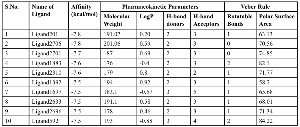
Table 1.1 Binding affinity, pharmacokinetic profile and Number of Rotatable bonds of top 10 ligands
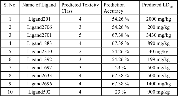
Table 1.2 Toxicity Profile of top 10 ligands obtained from ProTox-3.0
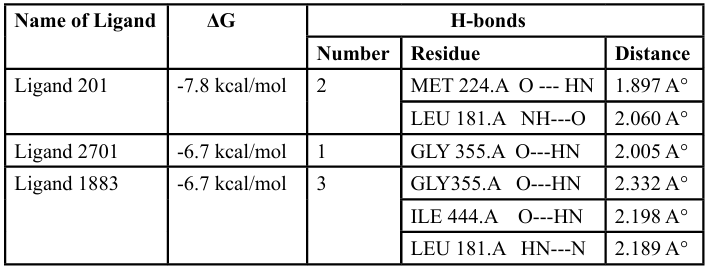
Table 1.3 Hydrogen Bonding of Ligand 201 with Receptor 3d11 in Autodock vina
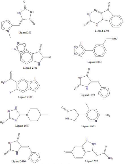
Figure 1. Top 10 ligands with binding affinity among 3,000 molecules
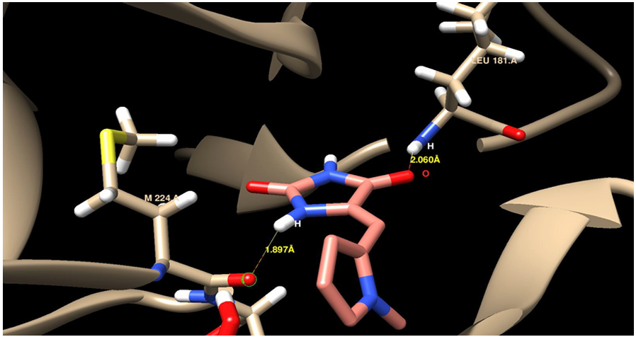
Figure 2. H-bonding of Ligand 201 with MET 224.A and LEU 181.A
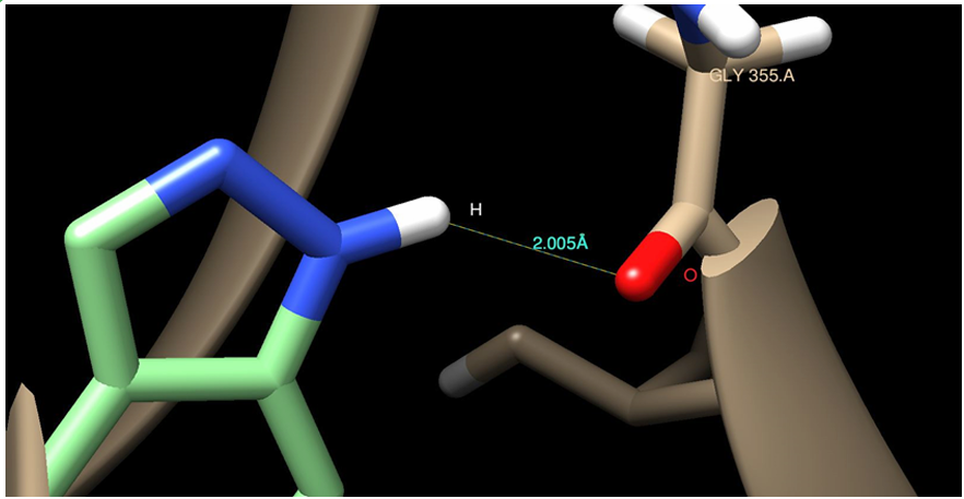
Figure 3. H-bonding of Ligand 2701 with GLY 355.A
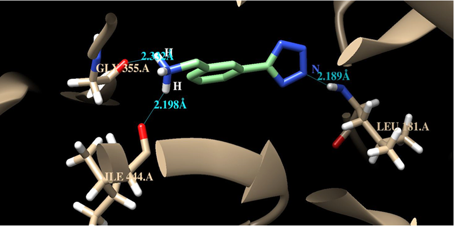
Figure 4. H-bonding of Ligand 2701 with GLY 355.A, ILE444.A and LEU181.A
Conclusion
The ongoing threat posed by the Nipah virus (NiV) necessitates urgent advancements in antiviral therapies, particularly given the its high mortality rate and lack of effective treatments. This study successfully employed virtual screening and molecular dynamics simulations to identify three promising inhibitors of the NiV glycoprotein (NiV-G)—Ligand201, Ligand2701, and Ligand1883. These compounds exhibited significant binding affinities, demonstrating potential to disrupt the interaction between NiV-G and its host cell receptors, thereby impeding viral entry.
The findings underscore the utility of computational methods in drug discovery, enabling the efficient evaluation of a large library of small molecules. This method has become a cornerstone in early drug discovery, enabling the identification of novel ligands based on protein structures [22]. The strong interactions of the identified ligands with key residues in the receptor-binding domain of NiV-G highlight their potential for further development. The favorable [18] pharmacokinetic profiles and low toxicity predicted for these compounds enhance their viability as lead candidates for subsequent in vitro and in vivo studies.
As future work aims to optimize these candidates for drug likeness and bioavailability, this research represents a critical step forward in addressing the public health challenges posed by Nipah virus outbreaks. The identification of effective inhibitors not only contributes to our understanding of NiV biology, but also sets a foundation for combating other emerging zoonotic viruses, reinforcing the importance of innovative therapeutic strategies in global health.
Conflict of interest:
The authors have no conflicts of interest to declare that are relevant to the content of this article.
References
Kerry, R. G., Malik, S., Redda, Y. T., Sahoo, S., Patra, J. K., & Majhi, S. (2019). Nano-based approach to combat emerging viral (NIPAH virus) infection. Nanomedicine: Nanotechnology, Biology and Medicine, 18, 196–220. View
Chua, K. B. (2003). Nipah virus outbreak in Malaysia. Journal of Clinical Virology, 26(3), 265–275. View
Chattu, V., Kumar, R., Kumary, S., Kajal, F., & David, J. (2018). Nipah virus epidemic in southern India and emphasizing “One Health” approach to ensure global health security. Journal of Family Medicine and Primary Care, 7(2), 275. View
Nipah virus infection. (n.d.). Retrieved September 14, (2024). View
Pandey, H., Pandey, P., Jakhmola, V., Semwal, A., Deorari, M., Ansori, A. N. M., & Zainul, R. (2024). A Comprehensive Review of Nipah Virus: From Epidemics to Approaches of Management. Journal of Pure and Applied Microbiology, 18(3), 1502–1514. View
Verma, M. K., Verma, P., Singh, S., Gaur, P., Siddiqui, A. H., & Pandey, S. (2018). Nipah Virus- Infectious Agent: An Overview. International Journal of Life-Sciences Scientific Research, 4(3), 1844–1850. View
Siddique, A. B., Fardows, J., Farhana, N., & Mazumder, M. (2016). Nipah Virus: A Public Health Concern. Journal of Enam Medical College, 6(2), 101–105. View
Nipah virus: Kerala state issues alert after 14-year-old dies. (n.d.). Retrieved September 14, 2024, from https://www.bbc. com/news/articles/cj50d7e9vp6o View
Health ministry alerts agencies to risk of Nipah virus outbreak. (n.d.). Retrieved September 14, 2024. View
Ortega, V., Zamora, J. L. R., Monreal, I. A., Hoffman, D. T., Ezzatpour, S., Johnston, G. P., Contreras, E. M., Vilchez Delgado, F. J., & Aguilar, H. C. (2022). Novel Roles of the Nipah Virus Attachment Glycoprotein and Its Mobility in Early and Late Membrane Fusion Steps. mBio, 13(3), e03222-21. View
Chan, X. H. S., Haeusler, I. L., Choy, B. J. K., Hassan, M. Z., Takata, J., Hurst, T. P., Jones, L. M., Loganathan, S., Harriss, E., Dunning, J., Tarning, J., Carroll, M. W., Horby, P. W., & Olliaro, P. L. (2024). Nipah Virus Therapeutics: A Systematic Review to Support Prioritisation for Clinical Trials. View
Rababi, D., & Nag, A. (2023). Evaluation of therapeutic potentials of selected phytochemicals against Nipah virus, a multi-dimensional in silico study. 3 Biotech, 13(6), 174. View
Negru, P. A., Miculas, D. C., Behl, T., Bungau, A. F., Marin, R.-C., & Bungau, S. G. (2022). Virtual screening of substances used in the treatment of SARS-CoV-2 infection and analysis of compounds with known action on structurally similar proteins from other viruses. Biomedicine & Pharmacotherapy, 153, 113432. View
Hollingsworth, S. A., & Dror, R. O. (2018). Molecular Dynamics Simulation for All. Neuron, 99(6), 1129–1143. View
Xu, K., Rajashankar, K. R., Chan, Y.-P., Himanen, J. P., Broder, C. C., & Nikolov, D. B. (2008). Host cell recognition by the henipaviruses: Crystal structures of the Nipah G attachment glycoprotein and its complex with ephrin-B3. Proceedings of the National Academy of Sciences, 105(29), 9953–9958. View
Ropón-Palacios, G., Chenet-Zuta, M. E., Olivos-Ramirez, G. E., Otazu, K., Acurio-Saavedra, J., & Camps, I. (2020). Potential novel inhibitors against emerging zoonotic pathogen Nipah virus: A virtual screening and molecular dynamics approach. Journal of Biomolecular Structure and Dynamics, 38(11), 3225–3234. View
Trott, O., & Olson, A. J. (2010). AutoDock Vina: Improving the speed and accuracy of docking with a new scoring function, efficient optimization, and multithreading. Journal of Computational Chemistry, 31(2), 455–461. View
Yang, Z., Ji, J., He, S., Li, J., He, T., Bai, R., Zhu, Z., & Ong, Y. S. (2024). Dockformer: A transformer-based molecular docking paradigm for large-scale virtual screening. View
Lipinski, C. A., Lombardo, F., Dominy, B. W., & Feeney, P. J. (2001). Experimental and computational approaches to estimate solubility and permeability in drug discovery and development settings 1PII of original article: S0169-409X(96)00423-1. The article was originally published in Advanced Drug Delivery Reviews 23 (1997) 3–25. 1. Advanced Drug Delivery Reviews, 46(1–3), 3–26. View
Veber, D. F., Johnson, S. R., Cheng, H.-Y., Smith, B. R., Ward, K. W., & Kopple, K. D. (2002). Molecular Properties That Influence the Oral Bioavailability of Drug Candidates. Journal of Medicinal Chemistry, 45(12), 2615–2623. View
Banerjee, P., Kemmler, E., Dunkel, M., & Preissner, R. (2024). ProTox 3.0: A webserver for the prediction of toxicity of chemicals. Nucleic Acids Research, 52(W1), W513–W520. View
Zhu, H., Zhang, Y., Li, W., & Huang, N. (2022). A comprehensive survey of prospective structure-based virtual screening for early drug discovery in the past fifteen years. International Journal of Molecular Sciences, 23(24), 15961. View