
- About the Journal
- Editorial Board
- Review Process
- Author Guidelines
- Article Processing Charges
- Special Issues
- Current Issue
- Past Issue
 Journal of Basic and Applied Pharmaceutical Science
 Volume 4 (2026), Article ID: JBAPS-112
https://doi.org/10.33790/jbaps1100112Mini Review
Optimizing Niosomes as Nanocarriers for Topical Antipsoriatic Therapy: A Mini-Review
María Julia Altube1, María José Morilla1, and Ana Paula Perez1*
1Nanomedicine Research and Development Centre, Science and Technology Department, National University of Quilmes, Roque Saenz Peña 352, Bernal, B1876, Buenos Aires, Argentina.
Corresponding Author Details: Ana Paula Perez, Nanomedicine Research & Development Center, Departamento de Cienciay Tecnología, Universidad Nacional de Quilmes. Roque Saenz Peña 352, Bernal, B1876, Buenos Aires, Argentina.
Received date: 10th November, 2025
Accepted date: 07th April, 2026
Published date: 09th April, 2026
Citation: Altube, M. J., Morilla, M. J., & Perez, A. P., (2026). Optimizing Niosomes as Nanocarriers for Topical Antipsoriatic Therapy: A Mini-Review. J Basic Appl Pharm Sci, 4(1): 112.
Copyright: ©2026, This is an open-access article distributed under the terms of the Creative Commons Attribution License 4.0, which permits unrestricted use, distribution, and reproduction in any medium, provided the original author and source are credited.
Abstract
Psoriasis is a chronic inflammatory autoimmune skin disorder characterized by keratinocyte hyperproliferation and immune dysregulation. Although topical therapy remains first-line for mild to moderate disease, conventional formulations are limited by poor drug penetration and potential adverse effects. Nanosized carriers, particularly niosomes, vesicular systems composed of non-ionic surfactants, offer a cost-effective and stable alternative capable of encapsulating hydrophilic and lipophilic drugs while enhancing skin retention and therapeutic efficacy. This mini-review summarizes eight studies from the past decade that employed Design of Experiments (DoE) approaches to optimize niosomal formulations for topical antipsoriatic therapy. Key formulation variables including surfactant type, surfactant: lipid ratio, hydration time and drug concentration were systematically adjusted to influence critical vesicle characteristics such as size and encapsulation efficiency. Optimized niosomes consistently improved drug deposition in the epidermis and viable dermis with low overall skin permeation, and in vivo models confirmed reductions in Psoriasis Area and Severity Index (PASI) scores and improvements in skin histology. These findings support niosomes as effective and rationally optimized nanocarriers for topical antipsoriatic therapy.
Introduction
Psoriasis
Psoriasis is a chronic, recurrent, inflammatory autoimmune skin disease affecting approximately 2–5% of the global population. Clinically, it manifests in forms such as plaque psoriasis, psoriatic arthritis, erythrodermic psoriasis, and pustular psoriasis. Among them, plaque psoriasis is the most prevalent, presenting as well- defined erythematous and scaly lesions. Beyond its cutaneous manifestations, psoriasis is associated with several comorbidities, including cardiometabolic disorders, psoriatic arthritis, and depression [1].
The etiology of psoriasis is multifactorial, arising from the interplay between genetic susceptibility, environmental triggers (such as streptococcal infections, stress, obesity, alcohol consumption, and smoking), and immune dysregulation. Disease development and progression are driven by T cell differentiation, infiltration of inflammatory cells, and hyperproliferation of keratinocytes. Histologically, psoriatic lesions display epidermal thickening, thinning of the granular layer, capillary dilation, and dense T cell infiltrates in both dermis and epidermis, often accompanied by neutrophil clusters in the stratum corneum. Pathogenesis results from a dynamic cross-talk between the innate and adaptive immune systems, mediated by cytokines such as TNF-α, IFN-γ, IL-17, and IL-22. These cytokines activate keratinocytes, promoting their hyperproliferation and the release of antimicrobial peptides, growth factors, and chemokines, which in turn drive angiogenesis, neutrophil infiltration, and the expansion of Th1 and Th17 populations. The outcome is a self-sustaining inflammatory cycle in which IL-23 and Th17 responses, together with TNF-α and interferons, play a central role [1,2].
The therapeutic goal in psoriasis is to control symptoms, reduce inflammation, and minimize relapse frequency. For mild to moderate plaques, topical formulations remain the first-line approach. These include corticosteroids, vitamin D analogs, calcineurin inhibitors, retinoids, keratolytic agents, and emollients, which act by modulating inflammation and keratinocyte proliferation and differentiation, while alleviating itching and scaling. However, topical therapies may induce adverse effects such as irritation, burning, pruritus, or local skin alterations, and in some cases, there is a risk of systemic absorption [3]. For moderate to severe forms, systemic therapies, biologics, or phototherapy are usually required. Although these options can be effective, they are associated with higher risks and limitations, and to date, there is no completely safe or definitive cure for the disease.
Despite their advantages, topical treatments face significant challenges. The penetration of drugs through the stratum corneum is often insufficient to reach therapeutic levels in thicker plaques or lesions located on the palms, soles, or scalp. To overcome these barriers, drug delivery nanosystems have emerged as a promising strategy for psoriasis therapy. Such systems enable controlled transport and release of active agents, enhancing their skin penetration and therapeutic efficacy while reducing adverse effects associated with systemic absorption [4]. Among these, lipid-based nanoparticles have been widely investigated due to their ability to improve drug stability, solubility, and bioavailability, promote wound healing and skin repair, and allow formulation with natural and biocompatible excipients [5]. However, while large-scale production of lipid-based nanoparticles can be challenging, this limitation is more critical for parenteral or pulmonary applications, whereas topical formulations may allow more feasible and cost-effective manufacturing [6,7].
Liposomes are the most extensively studied and developed lipid- based nanoparticles. In recent years, many liposomal derivatives have been designed to overcome some of the limitations of conventional liposomes [8,9]. Among these, niosomes are vesicular drug delivery systems composed of non-ionic surfactants. The replacement of phospholipids with these surfactants allows for the development of a less expensive and more stable nanocarrier [10]. Niosomes exhibit good chemical and physical stability, biocompatibility, and the ability to encapsulate both hydrophilic and hydrophobic drugs, making them an attractive option for topical drug delivery in psoriasis [11].
Niosomes
Niosomes were first reported in 1975 by researchers at L’Oréal, who patented them as vesicular systems intended for cosmetic applications. Since then, these nanocarriers have gained attention in diverse fields, including the pharmaceutical, cosmetic, and food industries [12]. Structurally, niosomes are vesicles composed of non-ionic surfactants that self-assemble into bilayers, resembling liposomes in both morphology and encapsulation mechanism (Figure 1). Like liposomes, they can entrap hydrophilic drugs within the aqueous core and incorporate lipophilic compounds into the bilayer [13].
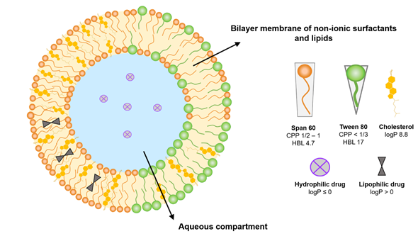
Figure 1. Schematic representation of a niosome structure. Niosomes are vesicular systems composed of a bilayer membrane formed by non-ionic surfactants (e.g., Span 60, Tween 80) and lipids, typically cholesterol, enclosing an aqueous compartment. The arrangement of amphiphilic molecules enables the encapsulation of both hydrophilic drugs within the aqueous core and lipophilic drugs within the bilayer membrane. The hydrophilic–lipophilic balance (HLB) and critical packing parameter (CPP) of the surfactants determine vesicle organization. Moreover, the surfactant-to-cholesterol ratio critically influences vesicle size, bilayer fluidity, encapsulation efficiency, and polydispersity.
Niosomes were originally developed to overcome several limitations associated with liposomes, particularly instability, high cost, and susceptibility to oxidation [14]. The non-ionic surfactants used in niosome formulation are amphiphilic molecules with a polar head and a non-polar tail, which play a similar structural role to phospholipids in liposomes but offer improved physicochemical stability and resistance to oxidative degradation [10]. Niosomes are typically formed from synthetic non-ionic surfactants with alkyl ether or ester chains. The formation of bilayers instead of micelles by non-ionic surfactants depends primarily on parameters such as the hydrophilic–lipophilic balance (HLB), which reflects the relative affinity of the polar head and hydrophobic tail for water and lipids, and the critical packing parameter (CPP), which predicts the preferred self-assembled structure according to the molecular geometry [15]. The HLB is an empirical measure of the relative proportion of hydrophilic and hydrophobic groups in a surfactant. Surfactants with higher HLB values are more water-soluble than those with lower HLB. This parameter is critical for niosome formation: surfactants with HLB values between 4 and 8, such as Span 40, 60, and 80, can spontaneously form stable niosomes. More hydrophilic surfactants, with HLB values of 8 or higher (e.g., Span 20, Tweens), generally require the addition of a co-surfactant or a lipid such as cholesterol to promote vesicle assembly, as excessive hydrophilicity can compromise structural stability. Surfactants with HLB values outside this range are typically unable to form stable niosomes [16].
The CPP is a dimensionless number that reflects the optimal geometric arrangement of amphiphilic molecules and the interactions between their polar and non-polar regions [17]. It provides insight into the limiting shapes that molecules can adopt and, consequently, the type of self-assembled structure they will form. CPP can be calculated using computational chemistry software or estimated from Tanford equations, which consider the number of carbons in the alkyl chain to determine the non-polar chain length, polar head area, and hydrophobic volume [15]. Mathematically, CPP is expressed as CPP = V / (a0 × lc), where V is the hydrophobic group volume, a0 is the area of the hydrophilic head, and lc is the critical hydrophobic group length. The CPP value predicts the resulting structure: values below 0.5 favor spherical micelles, whereas values between 0.5 and 1 promote the formation of bilayer structures such as niosomes [18]. In view of the above, it is important to select surfactants with suitable HLB and CPP values to favor bilayer formation, enabling the assembly of vesicular structures. Commonly used surfactants include members of the Span (20, 40, 60, 65, 80, 85), Tween (20, 40, 60, 65, 80, 85), and Brij (30, 35, 52, 56, 58) families, often combined with cholesterol to modulate membrane rigidity and permeability [18,19].
The phase transition temperature (Tc) of the surfactant also influences vesicle rigidity and stability. Surfactants with high Tc values, such as Span 60, form more rigid bilayers, enhancing encapsulation efficiency (EE%) and reducing membrane permeability, which minimizes premature drug release [20].
In addition to surfactant selection and physicochemical properties, the method of preparation plays a crucial role in determining niosome size, lamellarity, and EE% [15]. The thin film hydration method, also known as the stirring method, is the most commonly used approach. In this method, surfactants, stabilizers, and the drug are dissolved in an organic solvent, evaporated to form a thin film, and then hydrated to yield multilamellar vesicles containing the drug [21]. The solvent injection method involves slowly adding a surfactant–cholesterol mixture dissolved in an organic solvent to a preheated aqueous phase, followed by a phase transition to form vesicles, typically used for multilamellar niosomes encapsulating hydrophilic drugs [22]. Other techniques include the reverse evaporation phase method, where the organic phase is removed under reduced pressure after homogenization to entrap hydrophilic drugs [23], and the microfluidic hydrodynamic approach, which employs controlled diffusive mixing in microchannels to optimize vesicle size and polydispersity [24].
In addition to their favorable physicochemical properties, niosomes are particularly well suited for large-scale production. The higher chemical stability compared with liposomes allows them to maintain vesicle size, lamellarity, and structural integrity under industrial manufacturing conditions, which often involve variations in temperature, mixing rate, and processing time [10,12]. Synthetic non-ionic surfactants used in niosomes are more resistant to hydrolysis and oxidation than phospholipids, which minimizes aggregation and degradation during storage and scale-up [25]. Consequently, niosomes can be reliably produced in large quantities while preserving EE% and vesicle uniformity, making them attractive for industrial applications and translational research.
Optimization Analysis
Design of Experiments (DoE) is a key statistical tool in the development of pharmaceutical formulations, enabling the systematic optimization of processes and products through the simultaneous evaluation of multiple variables and their interactions. Unlike traditional trial-and-error or one-factor-at-a-time approaches, DoE provides a deeper scientific understanding of experimental systems while reducing the number of tests, development time, and associated costs [26]. This approach establishes clear relationships among formulation variables, process conditions, and final product attributes [27]. The implementation of DoE has proven particularly valuable in the design of vesicular drug delivery systems such as liposomes and niosomes, where small variations in formulation components or manufacturing conditions can significantly affect the stability of the vesicles, drug EE%, and bioavailability [28].
In modern pharmaceutical development, DoE is integrated within the Quality by Design (QbD) framework promoted by international regulatory agencies such as the U.S. Food and Drug Administration (FDA) and the International Council for Harmonisation (ICH). These initiatives encourage science- and risk-based approaches to ensure product quality from the earliest stages of development [29,30].
The present mini-review summarizes eight studies published over the past decade that employed DoE approaches to optimize niosomes for the topical delivery of antipsoriatic drugs. These studies explored how independent variables—such as surfactant-to-lipid ratio, surfactant type (HLB value), hydration time, and drug concentration— affect the physicochemical characteristics of niosomes, including vesicle size, polydispersity index (PDI), ζ potential, and EE%. Although not all studies measured the same parameters, this mini review focuses on vesicle size and EE%, which were consistently reported and allow cross-study comparison. In addition, findings on skin penetration and in vivo performance are discussed. The key methodological and formulation characteristics of the reviewed studies are compiled in Table 1.
Optimization of Niosomes for the Topical Delivery of Antipsoriatic Drugs
Although the mechanisms by which niosomes could enhance drug deposition in the epidermis and viable dermis remain poorly validated experimentally, they are considered promising for efficient delivery of antipsoriatic drugs. It has been suggested that the surfactant components of niosomes may act as permeation enhancers and that niosomes can reduce transepidermal water loss, thereby increasing skin hydration and facilitating drug accumulation [31]. Studies on liposomes have further proposed that vesicle properties such as size and surface charge influence skin penetration: vesicles larger than 600 nm tend to remain in the stratum corneum, those below 300 nm can reach deeper layers to a limited extent, and vesicles under 70 nm may significantly accumulate in deeper tissues [32]. Additionally, negatively charged vesicles have been reported to diffuse faster through the dermis and lower hair follicle regions compared to positively charged vesicles, which may further contribute to enhanced local deposition [33].
Across the reviewed studies, optimization efforts consistently aimed to obtain niosomes with sizes generally in the 100–500 nm range, proposed to promote deposition within the stratum corneum and viable epidermis while minimizing systemic absorption. However, one study reported larger vesicles (~1345 nm) as optimal for maximizing skin retention and minimizing transdermal permeation [34]. Uniformity of vesicle size is commonly assessed using the PDI, with low polydispersity (PDI < 0.3) ensuring a consistent vesicle population and reproducible topical performance. High EE% increases the amount of drug available for sustained localization in the skin, whereas absolute ζ potential values above 30 mV contribute to colloidal stability and minimize aggregation during storage and application.
Together, these parameters define a performance window considered optimal for maximizing dermal delivery and therapeutic efficacy of niosomal formulations for psoriasis in the reviewed studies.
Effects on Niosome Size
The size of niosomes primarily depends on the balance between the surfactant and cholesterol, which determines bilayer rigidity, curvature, and packing density. The magnitude and trend of this effect vary according to the surfactant nature and its HLB value.
In formulations prepared with Span 60 (HLB ≈ 4.7, indicating a predominantly lipophilic character), an increase in surfactant concentration was associated with a decrease in vesicle size, attributed to greater bilayer curvature and the formation of more compact vesicles with lower polydispersity [28,35]. This behavior can be explained by the low CPP of Span 60, which favors the formation of more tightly closed structures [36]. Consistently, a decrease in the Span 60: cholesterol ratio resulted in an increase in niosome size, due to the intercalation of cholesterol among the hydrocarbon chains of the surfactant, which enhances bilayer order and rigidity. This structural reinforcement reduced deformability and limits size reduction during sonication or stirring processes [37]. The high lipophilicity of cholesterol (log P ≈ 7) further contributes to lower membrane flexibility and the formation of larger, more stable vesicles. According to Pandey et al., a slight increase in lipid content within the Span 60: cholesterol ratio also tended to increase vesicle diameter, probably due to bilayer thickening, although this effect was not statistically significant [38].
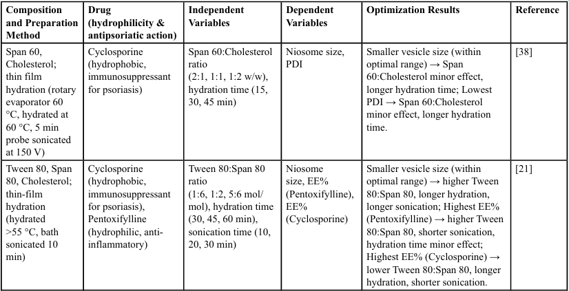
Table 1. Optimization of niosomes for topical antipsoriatic drug delivery
Similarly, Jafarian et al., employing Span 60 and Tween 80 as surfactants and 1-dodecanol as a co-surfactant/stabilizer in tretinoin loaded formulations, observed that an increase in the surfactant:1 dodecanol ratio significantly reduced niosome size [39]. This effect was attributed to higher bilayer cohesion and compaction, resulting in smaller vesicles with lower polydispersity.
Conversely, Abdelbary et al. reported that increasing the Span 60: cholesterol ratio led to larger vesicles in methotrexate-loaded formulations [34]. The authors attributed this finding to a higher drug-loading capacity within the aqueous core, resulting in vesicles of greater volume. A similar behavior was described by Bhardwaj et al. in cyclosporine-loaded niosomes, where higher Span 60 content correlated with increased vesicle size, accompanied by a simultaneous rise in the EE% of the lipophilic drug [40].
In Tween 80-based formulations (HLB ≈ 15, indicating a highly hydrophilic surfactant), Bhardwaj et al. observed that increasing the surfactant-to-cholesterol ratio produced larger vesicles [41]. This effect was linked to greater water uptake by the bilayer and the resulting increase in system fluidity, which promoted vesicle expansion.
Taken together, the relative and absolute content of cholesterol and surfactant exert a decisive influence on niosome size. In general, cholesterol increases vesicle size by enhancing membrane rigidity, whereas lipophilic surfactants (e.g., Span 60) tend to yield smaller and more compact vesicles, while hydrophilic surfactants (e.g., Tween 80) favor the formation of larger and more flexible structures. It is worth noting that vesicle flexibility influences skin transport processes [42].
The hydration time of the film is another critical parameter that determines how surfactant and cholesterol molecules organize into stacked bilayers and the extent to which these layers expand and close to form stable vesicular assemblies, ultimately influencing the final niosome size. In most of the reviewed studies, a longer hydration process of 45-60 minutes was associated with a significant reduction in vesicle size. This effect has been attributed to a more complete reorganization of the surfactant/cholesterol bilayer, allowing molecules to arrange into more ordered continuous structures, and to the disruption of multilamellar vesicles into smaller and more uniform vesicles.
For instance, Pandey et al., reported that a 15-minute increase in hydration time reduced the average niosome size by approximately 64 nm, an effect explained by enhanced swelling of the lipid film, which facilitates the release of interlamellar tension and promotes the formation of more compact vesicles [38]. Similarly, Bhardwaj et al. observed a progressive decrease in vesicle size when the hydration time was extended from 30 to 60 minutes, attributed to the disruption of larger vesicles and reorganization of the colloidal system [21]. Consistent results were observed by Bhardwaj et al. in cyclosporine- loaded formulations, where prolonged hydration (60 minutes) led to smaller niosomes [40]. The authors suggested that extended hydration promotes swelling of the surfactant/cholesterol bilayer, increasing fluidity and loosening the packing of surfactant/lipid layers, leading to the disruption of larger multilamellar vesicles and the formation of smaller unilamellar populations. In contrast, very short hydration periods may result in partially hydrated films with incomplete bilayer formation, yielding vesicles with larger diameters or heterogeneous size distribution. A similar pattern was described by Shah et al., where extended mixing and hydration times contributed to the formation of smaller and more uniform vesicles [28]. In line with these observations, Moghddam et al. reported a linear decrease in vesicle size with increasing hydration time, which they attributed to disruption of the vesicular structure and drug leakage at longer hydration durations [35].
These findings suggest a trend toward a decrease in vesicle size with increasing hydration time.
Effects on EE%
EE% is a key parameter because it determines how much of the drug is initially incorporated into the vesicles, which directly influences the amount available for localized and sustained delivery within the skin. Upon topical application, niosomes act as transient reservoirs that enhance skin penetration and promote controlled drug accumulation in epidermal and dermal layers [43]. This targeted deposition increases drug availability within localized skin layers while reducing the required dose and minimizing systemic exposure. Consequently, higher EE% supports more efficient dermal delivery and a smoother release pattern, ultimately improving efficacy and tolerability. Several studies have analyzed how formulation variables affect EE% in niosomes, showing that these effects strongly depend on the drug properties and the niosome composition.
Cholesterol plays a dual role in drug encapsulation. In formulations containing hydrophilic drugs such as pentoxifylline, increasing cholesterol content enhanced EE% by stiffening the bilayer and decreasing its permeability [41]. However, for other hydrophilic drugs, such as methotrexate, decreasing the cholesterol content significantly increased EE%, likely because higher cholesterol levels can disrupt the regular structure of the vesicle membrane, limiting drug encapsulation [34]. In formulations with lipophilic drugs such as cyclosporine [40] and diacerein [35], excessive cholesterol decreased EE%, likely due to competition with the drug within the lipid bilayer. Overall, a moderate cholesterol content appears optimal, improving stability without compromising loading capacity.
Although the reviewed studies did not explicitly discuss the influence of surfactant HLB on EE%, this concept helps interpret the observed trends related to surfactant concentration. Surfactants with low HLB values (e.g., Span 60 or 80) exhibit higher affinity for lipid phases and promote compact bilayer formation, whereas those with high HLB values (e.g., Tween 80) are more water- soluble and increase vesicle flexibility. In the analyzed formulations, Bhardwaj et al. observed that increasing the Tween 80: Span 80 ratio i.e., increasing the fraction of the higher-HLB surfactant enhanced EE% for the hydrophilic drug pentoxifylline but decreased it for the lipophilic cyclosporine [21]. Consistently, Moghddam et al. reported that increasing Span 60, with lower HLB, improved the encapsulation of diacerein (lipophilic) [35], while Jafarian et al. demonstrated that higher surfactant concentrations, independent of HLB, led to smaller vesicles, and since drug-loading efficiency may be correlated with particle size, this resulted in lower encapsulation of hydrophobic drugs [39]. These findings suggest that the relationship between surfactant HLB and drug polarity can guide formulation design, although the final outcome depends on multiple factors such as surfactant content and vesicle size.
Among the reviewed studies, hydration time mainly affected formulations containing lipophilic drugs. Bhardwaj et al. reported that prolonged hydration increased EE% for cyclosporine, probably due to more complete incorporation into the lipid bilayer [40]. In contrast, Moghddam et al. observed a decrease in EE% for diacerein at extended hydration times, possibly due to vesicle reorganization or destabilization [35]. Similarly, Shah et al., using the ether injection method, showed that longer mixing times and lower agitation speeds increased EE% by promoting the formation of more compact and stable vesicles [28].
Regarding drug amount, the effects on EE% depend on how this variable is modified. Bhardwaj et al. increased the mass of pentoxifylline while presumably keeping the hydration volume constant, observing a decrease in EE%; the authors attributed this to an increase in the “effective hydration volume” [41]. Conversely, Abdelbary et al. maintained the total methotrexate mass but reduced the hydration volume, which increased EE% by concentrating the drug within the medium [34]. These results indicate that the effect of drug amount critically depends on whether the total mass or the effective concentration in the hydration medium is altered. On the other hand, Bhardwaj et al. observed that EE% of cyclosporine increased with higher drug amounts, probably due to saturation conditions that favored greater incorporation of the active compound into the niosomes [40].
In summary, the DoE-based optimization processes applied in the reviewed studies proved robust and predictive. In all cases, the close agreement between predicted and experimental responses validated the optimization approach and supported the selection of variable combinations that enhanced the targeted niosome attributes.
Biological Results of the Optimized Formulations
The biological performance of the optimized niosome formulations was assessed by studying skin penetration in ex vivo and in vivo models, as well as their antipsoriatic effects in animal models of psoriasis, compared with the free drug. The results are summarized in Table 2.
Table 2. Physicochemical characteristics and biological performance of optimized niosomes for topical psoriasis therapy
Evaluation of Skin Penetration
Most of the reviewed studies employed Franz diffusion cells to evaluate ex vivo skin permeation and deposition, using skin from different species, including albino rats, goats, and human cadaver skin, which makes direct comparison between studies challenging. This methodology allows the determination of both drug permeation across the skin and deposition within the epidermis and viable dermis layers. In the context of localized topical therapy, however, the primary goal is not high permeation but rather efficient accumulation within the target skin layers. It should be noted that these models involve healthy skin and do not reproduce the altered barrier properties of psoriatic skin.
Pandey et al. conducted ex vivo studies on albino rat abdominal skin and observed that cyclosporine suspension deposited only 0.39 % and 0.59 % in the stratum corneum and viable epidermis/dermis, respectively, with most of the drug (88.8 %) remaining on the skin surface [38]. In contrast, cyclosporine-loaded niosomes markedly increased deposition (15.1 % and 42.7 %), while incorporation into a Carbopol 940 gel slightly reduced deposition (13 % and 30 %) but still outperformed the free drug. Permeation across the skin was higher for niosomes (51 %) compared to suspension (10 %), and slightly lower when incorporated into the gel (31 %) [38].
Similar trends were observed by Bhardwaj et al. in goat skin, where cyclosporine-loaded niosomes increased deposition in the stratum corneum and viable epidermis/dermis (~15.8 % and ~36 %) compared to the drug suspension (3.2 % and 7.3 %) after 10 hours, while the amount permeated across the skin remained higher for niosomes (13 % vs. 6.2 %) [40].
Bhardwaj et al. further demonstrated that pentoxifylline exhibited low permeation when delivered alone, but co-loading with cyclosporine in niosomes significantly enhanced deposition in the stratum corneum and viable dermis/epidermis layers [21]. After 24 h, deposition from niosomes was markedly higher than from the free drugs, while permeation remained relatively low (3.4 % for pentoxifylline and 9.9 % for cyclosporine vs. 0.6 % and 6.2 % for the free drugs, respectively). In another study on goat skin, deposition of pentoxifylline from niosomes reached 11 % in the stratum corneum and 31 % in the viable epidermis/dermis, compared to 0.8 % and 2.3 % for the solution formulation [41].
Shah et al. evaluated desoximetasone niosomal gel on dermatomed human cadaver skin [44]. Although the flux of the reference gel was higher, the niosomal gel tended to retain more drug within the skin (31 ng/mg vs. 20 ng/mg), although the difference was not statistically significant.
Moghddam et al. investigated diacerein niosomes in albino rat skin [35]. The highest flux observed was 2.82 μg/cm²/h, and confocal laser scanning microscopy of Rhodamine B-labeled niosomes revealed penetration up to 180 μm, with the greatest intensity between 30 and 90 μm, corresponding to the epidermal layer.
Abdelbary et al. performed an in vivo skin deposition study in Wistar rats of a methotrexate loaded niosome formulation aimed at topical management of psoriasis [34]; deposition in skin was 22.5 % for the niosomes versus 13.9 % for the drug solution, illustrating a clear enhancement of localized skin retention.
Taken together, these studies show that niosomal formulations consistently enhanced local drug deposition in the epidermis and viable dermis layers compared to free drug solutions or suspensions, highlighting their potential for topical localized therapy, while permeation through the full skin thickness remained generally low. However, evidence for clear structure–activity relationships is still limited, and extrapolation of these findings to psoriatic skin should be made with caution. Some of the reviewed works also include in vivo evaluations, which are discussed separately below.
In Vivo Studies in a Psoriatic Mouse Model
Among the selected studies, those that performed in vivo assays employed the imiquimod-induced psoriatic plaque model in Swiss mice. In this model, a 5 % (w/w) imiquimod cream (approximately 62.5 mg) is applied daily to the shaved dorsal skin for seven consecutive days to induce psoriasis-like lesions. From day 7 to day 14, animals receive the corresponding treatments, and disease severity is assessed using the Psoriasis Area and Severity Index (PASI). The PASI score considers erythema, scaling, and skin thickness, each graded on a 5-point scale (0 = none, 1 = mild, 2 = moderate, 3 = severe, and 4 = very severe) by a trained evaluator. Histopathological analysis of skin samples is also conducted to corroborate clinical observations.
Pandey et al. reported that animals treated with a gel containing cyclosporine loaded in niosomes showed a significant reduction in PASI scores compared with both the untreated group and the group treated with a cyclosporine suspension [38]. Histological analysis revealed a reduction in epidermal thickness and stratum corneum in the niosome-treated group, whereas the cyclosporine suspension group retained typical features of psoriatic skin.
Bhardwaj et al. reported that niosomal pentoxifylline markedly reduced inflammation compared with both untreated controls and non-niosomal formulations [41]. In a subsequent study, the same group evaluated niosomes co-encapsulating cyclosporine and pentoxifylline observing a pronounced therapeutic improvement: the group treated with the co-loaded niosomes exhibited a marked reduction in PASI scores, decreasing from 4 on day 7 to 1 on day 14, indicative of near-complete recovery of psoriatic lesions [21]. Histopathological analysis revealed that niosomes containing only pentoxifylline slightly improved the stratum corneum and keratin layers, niosomes containing only cyclosporine improved only the keratin layer, and co-encapsulation of both drugs produced the most pronounced effect, reducing the thickness of both the epidermis and stratum corneum. Bhardwaj et al. confirmed these findings, demonstrating that animals treated with cyclosporine in suspension exhibited PASI scores similar to untreated controls, whereas the cyclosporine niosome-treated group showed a significant reduction in scores from four to less than one over seven days of treatment [40]. These effects were accompanied by improvements in both the stratum corneum and keratin layers.
Considering all results, these studies highlight the enhanced therapeutic efficacy of niosome formulations, particularly those combining multiple active compounds, in reducing psoriatic severity and restoring skin histology in the imiquimod-induced mouse model.
Conclusion
The studies reviewed demonstrate that careful optimization of niosome formulations through control of surfactant type and ratio, cholesterol content, hydration time, and drug concentration directly influences vesicle size and EE%, which are critical for effective topical delivery. Optimized niosomes consistently achieved sizes typically within the submicron range (≈100–500 nm), although larger vesicles were also reported, with low PDI, and high EE%. These properties translated into enhanced drug penetration and retention in the epidermis and viable dermis. In vivo studies using imiquimod- induced psoriatic mouse models confirmed that the optimized niosome formulations led to significant reductions in PASI scores and restoration of epidermal and stratum corneum morphology. Taken together, optimized niosomes showed improved drug delivery and therapeutic outcomes in psoriasis.
Conflict of interest:
The authors declare no conflicts of interest relevant to the content of this article.
Acknowledgments
MJA, MJM and APP are members of the Research Career Program from National Council for Scientific and Technological Research (CONICET), Argentina
Declaration of generative AI in scientific writing
The authors declare that ChatGPT was used to assist in rephrasing certain sentences. All content was thoroughly reviewed and edited by the authors, who accept full responsibility for the accuracy and integrity of the published article.
References
Griffiths, C. E. M., Armstrong, A. W., Gudjonsson, J. E., & Barker, J. N. W. N. (2021). Psoriasis. The Lancet, 397:1301–15. View
Sieminska, I., Pieniawska, M., & Grzywa, T. M. (2024). The Immunology of Psoriasis Current Concepts in Pathogenesis. Clin Rev Allergy Immunol, 66:164–91. View
Elmets, C. A., Korman, N. J., Prater, E. F., Wong, E. B., Rupani, R. N., & Kivelevitch, D., et al. (2021). Joint AAD–NPF Guidelines of care for the management and treatment of psoriasis with topical therapy and alternative medicine modalities for psoriasis severity measures. J Am Acad Dermatol, 84:432–70. View
Bodnár, K., Fehér, P., Ujhelyi, Z., Bácskay, I., & Józsa, L., (2024). Recent Approaches for the Topical Treatment of Psoriasis Using Nanoparticles. Pharmaceutics, 16:449. View
Perez, A. P., Altube, M. J., Schilrreff, P., Apezteguia, G., Celes, F. S., & Zacchino, S., et al. (2016). Topical amphotericin B in ultradeformable liposomes: Formulation, skin penetration study, antifungal and antileishmanial activity in vitro. Colloids Surf B Biointerface, 139:190–8. View
Aziz Hazari, S., Kaur, H., Karwasra, R., Abourehab, M. A. S., Ali Khan, A., & Kesharwani, P. (2023). An overview of topical lipid-based and polymer-based nanocarriers for treatment of psoriasis. Int J Pharm, 638:122938. View
Simioni, Y. R., Ricatti, F., Salvay, A. G., Jerez, H. E., Schilrreff, P., & Romero, E. L., et al. (2024). Activity of hydrogel-vitamin D3 /bacterioruberin nanoparticles on imiquimod-induced fibroblasts-keratinocytes spheroids. J Drug Deliv Sci Technol, 97:105738. View
Bartelds, R., Nematollahi, M. H., Pols, T., Stuart, M. C. A., Pardakhty, A., & Asadikaram, G., et al. (2018). Niosomes, an alternative for liposomal delivery. PLoS One, 13:e0194179. View
Romero, E. L., & Morilla, M. J., (2022). Nanoarchaeosomes in Drug Delivery, p. 149–86. View
Izhar, M. P., Hafeez, A., Kushwaha, P., & Simrah. (2023). Drug Delivery Through Niosomes: A Comprehensive Review with Therapeutic Applications. J Clust Sci, 34:2257–73. View
Monohar, S., Malarvannan, M., Dineshkumar, B., Prajapati, B., & Paul, D. (2025). Liposomes and Niosomes in Psoriasis Therapy: Emerging Vesicular Approaches for Targeted Treatment. Biomedical Materials & Devices. View
Liga, S., Paul, C., Moacă, E-A., & Péter, F., (2024). Niosomes: Composition, Formulation Techniques, and Recent Progress as Delivery Systems in Cancer Therapy. Pharmaceutics, 16:223. View
Baillie, A. J., Florence, A. T., Hume, L. R., Muirhead, G. T., & Rogerson, A. (1985). The preparation and properties of niosomes non-ionic surfactant vesicles. Journal of Pharmacy and Pharmacology, 37:863–8. View
El Maghraby, G., & Williams, A. (2009). Vesicular systems for delivering conventional small organic molecules and larger macromolecules to and through human skin. Expert Opin Drug Deliv, 6:149–63. View
Bautista-Solano, A. A., Dávila-Ortiz, G., Perea-Flores, M. J., & Martínez-Ayala, A. L. (2025). A Comprehensive Review of Niosomes: Composition, Structure, Formation, Characterization, and Applications in Bioactive Molecule Delivery Systems. Molecules, 30:3467. View
Khoee, S., & Yaghoobian, M. (2017). Niosomes: a novel approach in modern drug delivery systems. Nanostructures for Drug Delivery, Elsevier, p. 207–37. View
Israelachvili, J. N., Marčelja, S., & Horn, R. G. (1980). Physical principles of membrane organization. Q Rev Biophys, 13:121- 200. View
Kumar, G. P., & Rajeshwarrao, P. (2011). Nonionic surfactant vesicular systems for effective drug delivery an overview. Acta Pharm Sin B, 1:208–19. View
Böttger, R., Pauli, G., Chao, P-H., AL Fayez, N., Hohenwarter, L., & Li, S-D., (2020). Lipid-based nanoparticle technologies for liver targeting. Adv Drug Deliv Rev, 154–155:79–101. View
Witika, B. A., Bassey, K. E., Demana, P. H., Siwe-Noundou, X., & Poka, M. S. (2022). Current Advances in Specialised Niosomal Drug Delivery: Manufacture, Characterization and Drug Delivery Applications. Int J Mol Sci, 23:9668. View
Bhardwaj, P., Tripathi, P., Pandey, S., Gupta, R., & Ramchandra Patil, P. (2022). Cyclosporine and Pentoxifylline laden tailored niosomes for the effective management of psoriasis: In-vitro optimization, Ex-vivo and animal study. Int J Pharm, 626:122143. View
Moghassemi, S., Hadjizadeh, A., & Omidfar, K. (2017). Formulation and Characterization of Bovine Serum Albumin Loaded Niosome. AAPS PharmSciTech, 18:27–33. View
Karim, K., Mandal, A., Biswas, N., Guha, A., Chatterjee, S., & Behera, M., et al. (2010). Niosome: A future of targeted drug delivery systems. J Adv Pharm Technol Res, 1:374. View
Khatoon, M., Shah, K. U., Din, F. U., Shah, S. U., Rehman, A. U., & Dilawar, N., et al. (2017). Proniosomes derived niosomes: recent advancements in drug delivery and targeting. Drug Deliv, 24:56–69. View
Kodel, H. de A. C., Alizadeh, P., Ebrahimi, S. N., Machado, T. O. X., Oliveira, M. B.P. P., & Fathi F, et al. (2025). Liposomes and Niosomes: New trends and applications in the delivery of bioactive agents for cancer therapy. Int J Pharm, 668:124994. View
Frey, D. D., Engelhardt, F., & Greitzer, E. M. (2003). A role for “one-factor-at-a-time” experimentation in parameter design. Res Eng Des, 14:65–74. View
Yu, L. X., Amidon, G., Khan, M. A., Hoag, S. W., Polli, J., & Raju, G. K., et al. (2014). Understanding Pharmaceutical Quality by Design. AAPS J, 16:771–83. View
Shah, P., Goodyear, B., Haq, A., Puri, V., & Michniak-Kohn, B. (2020). Quality by Design: A systematic approach using an advanced statistical tool for optimization of niosomes for topical delivery. World Journal Of Pharmacy And Pharmaceutical Sciences SJIF, 632:251–77. View
Zhang, L., & Mao, S. (2017). Application of quality by design in the current drug development. Asian J Pharm Sci, 12:1–8. View
Yu, L. X. (2008). Pharmaceutical Quality by Design: Product and Process Development, Understanding, and Control. Pharm Res, 25:781–91. View
Zeb, A., Arif, S. T., Malik, M., Shah, F. A., Din, F. U., & Qureshi, O. S., et al. (2019). Potential of nanoparticulate carriers for improved drug delivery via skin. J Pharm Investig, 49:485–517. View
Verma, D. (2003). Particle size of liposomes influences dermal delivery of substances into skin. Int J Pharm, 258:141–51. View
Ogiso, T., Yamaguchi, T., Iwaki, M., Tanino, T., & Miyake, Y. (2001). Effect of Positively and Negatively Charged Liposomes on Skin Permeation of Drugs. J Drug Target, 9:49–59. View
Abdelbary, A. A., & Aboughaly, M. H. H. (2015). Design and optimization of topical methotrexate loaded niosomes for enhanced management of psoriasis: Application of Box Behnken design, in-vitro evaluation and in-vivo skin deposition study. Int J Pharm, 485:235–43. View
Moghddam, S. R. M., Ahad, A., Aqil, M., Imam, S. S., & Sultana, Y. (2016). Formulation and optimization of niosomes for topical diacerein delivery using 3-factor, 3-level Box-Behnken design for the management of psoriasis. Materials Science and Engineering C, 69:789–97. View
Soni, S., Baghel, K., Soni, M. L., Kashaw, S. K., & Soni, V. (2024). Size-dependent effects of niosomes on the penetration of methotrexate in skin layers. Futur J Pharm Sci, 10:48. View
Nasseri, B. (2005). Effect of cholesterol and temperature on the elastic properties of niosomal membranes. Int J Pharm, 300:95- 101. View
Pandey, S. S., Shah, K. M., Maulvi, F. A., Desai, D. T., Gupta, A. R., & Joshi, S. V., et al. (2021). Topical delivery of cyclosporine loaded tailored niosomal nanocarriers for improved skin penetration and deposition in psoriasis: Optimization, ex vivo and animal studies. J Drug Deliv Sci Technol, 63:102441. View
Jafarian, A., Khalilabadi, S., Shadloo, A., & Peyvandi, K. (2025). The Influence of Operational Parameters on the Characterization of Tretinoin-Loaded Niosomes. ACS Omega, 10:30031–45. View
Bhardwaj, P., Tripathi, P., Pandey, S., Chaurasia, D., & Ramchandra Patil, P. (2023). Tailored Non-ionic Surfactant Vesicles of Cyclosporine for the Treatment of Psoriasis: Formulation, Ex-Vivo and In-Vivo investigation-Application of Box-Behnken Design. Advanced Materials Science and Technology, 5:0–0. View
Bhardwaj, P., Tripathi, P., Pandey, S., Gupta, R., Khar, R. K., & Patil, P. R. (2022). Improved dermal delivery of pentoxifylline niosomes for the management of psoriasis: Development, optimization and in-vivo studies in imiquimod induced psoriatic plaque model. J Drug Deliv Sci Technol, 75. View
Peralta, M. F., Guzmán, M. L., Pérez, A. P., Apezteguia, G. A., Fórmica, M. L., & Romero, E. L., et al. (2018). Liposomes can both enhance or reduce drugs penetration through the skin. Sci Rep, 8:13253. View
Singh, S., Sharma, N., Behl, T., Sarkar, B. C., Saha, H. R., & Garg, K., et al. (2021). Promising Strategies of Colloidal Drug Delivery-Based Approaches in Psoriasis Management. Pharmaceutics, 13:1978. View
Shah, P., Goodyear, B., Dholaria, N., Puri, V., & Michniak Kohn, B.. (2021). Nanostructured non-ionic surfactant carrier based gel for topical delivery of desoximetasone. Int J Mol Sci, 22:1–27. View