
- About the Journal
- Editorial Board
- Review Process
- Author Guidelines
- Article Processing Charges
- Special Issues
- Current Issue
- Past Issue
 Journal of Rehabilitation Practices and Research
Journal of Rehabilitation Practices and Research Volume 5 (2024), Article ID: JRPR-148
https://doi.org/10.33790/jrpr1100148Case Report
Objective Evaluation of Local Muscle Fatigue in Cerebral Palsy Using A Gait Corrector
Masaharu Morita1*, Masafumi Katayama2
1 Department of Physical Therapy, Faculty of Medical Science, Fukuoka International University Health and Welfare, 3-6-40 Momochihama, Sawaraku, Fukuoka, 814-0001, Japan.
2 Department of Medical Laboratory Sciences, Faculty of Health Sciences, Junshin Gakuen University, 1-1-1 Chikushigaoka, Minamiku, Fukuoka, 815-8510, Japan.
Corresponding Author Details: Masaharu Morita, Professor, Department of Physical Therapy, Faculty of Medical Science, Fukuoka International University Health and Welfare, 3-6-40 Momochihama, Sawaraku, Fukuoka, 814-0001, Japan.
Received date: 30th December, 2023
Accepted date: 03rd February, 2024
Published date: 05th February, 2024
Citation: Morita, M., & Katayama, M., (2024). Objective Evaluation of Local Muscle Fatigue in Cerebral Palsy Using A Gait Corrector. J Rehab Pract Res, 5(1):148.
Copyright:©2024, This is an open-access article distributed under the terms of the Creative Commons Attribution License 4.0, which permits unrestricted use, distribution, and reproduction in any medium, provided the original author and source are credited.Creative Commons Attribution License, which permits unrestricted use, distribution, and reproduction in any medium, provided the original author and source are credited
Abstract
The participants were seven individuals (age range 7–36 years) whose walking ability ranged from independent to requiring assistance with a cane, walker, etc. The protocol consisted of resting for at least five minutes before starting to walk, wearing a Gait Corrector device, and walking with light and heavy loads based on the participant's rating of perceived exertion (RPE), followed by resting for 10 minutes before the end of the protocol.
Near-infrared spectroscopy (NIRS) electrodes were attached bilaterally to the tibialis anterior and lateral gastrocnemius muscles. NIRS was used to measure blood oxygenation dynamics in local muscles from rest before walking to 10 minutes after walking, and evoked electromyography was used to measure the nerve conduction velocity of the peripheral nerves before and during early and late walking.
The tone state of the lateral gastrocnemius muscle was measured three times using an evoked potential-testing device: before walking, immediately after heavy-load walking, and 10 minutes after walking. A recording electrode was attached to the abductor digitorum medialis muscle and a reference electrode was attached to the base of the toe. The M and F waves were recorded using tibial nerve stimulation. The average F/M ratio was used as a parameter of the F-wave.
Regarding the change in muscle tone, four of the seven patients exhibited no change or a lower mean F/M ratio after walking than before walking, whereas three patients who exhibited resistance to other dorsiflexion movements of the ankle joint had higher values. Blood oxygenation dynamics revealed a greater disparity between oxyhemoglobin and deoxyhemoglobin concentrations after walking in all participants, suggesting local muscle fatigue, the degree of which showed no improvement even after 10 minutes of rest.
Keywords: Fatigue of Local Muscle, Blood Oxygenation Dynamics, Evoked Electromyogram, Nerve Conduction Velocity
Introduction
In this study, a Gait Corrector device (a short leg brace with a ling bar; Hart Walker Japan Co. Ltd.) was applied to individuals with cerebral palsy, and the relationship between muscle tone during walking and local muscle fatigue was quantitatively analyzed using the nerve conduction velocity of evoked electromyograms and near infrared spectroscopy (NIRS). Abnormal muscle tone during walking tends to increase with movement execution. Moreover, it is important to measure and analyze the degree of local muscle fatigue within the range of daily walking to confirm the effectiveness of gait corrector application.
For many children with cerebral palsy who are forced to use wheelchairs, canes, or walkers with upper-extremity support, there has been active development and marketing of wheelchairs and walkers with upper-extremity support. There has, however, been less development of orthotics to help them stand and walk safely.
Although there have been reports of improved range of motion of the joints and the effects of suppression of lower extremity muscle tone in children with cerebral palsy [1,2], there have been few reports to date on the suppression of internal and external rotation of the hip joint and clarification of the relationship between muscle tone and local muscle fatigue.
This study aimed to apply a Gait Corrector device to individuals with cerebral palsy and hypertonia in the lower extremities and to noninvasively and quantitatively analyze the state of local muscle fatigue and muscle tone in walking before and after the application of NIRS and nerve conduction velocity in evoked electromyograms to determine the relationship between local muscle fatigue and muscle tone.
As a quantitative analysis to clarify the clinical significance of a Gait Corrector device based on previous basic research and as a part of NIRS, we employed the blood oxygen dynamics (difference in concentration change between oxyhemoglobin and deoxyhemoglobin) of the local muscles involved in ankle plantar dorsiflexion (lateral gastrocnemius and tibialis anterior muscles) as a measure of muscle fatigue, measured up to 10 minutes after walking. Furthermore, the degree of spastic muscle tone before and after walking was confirmed using the maximum amplitude ratio of the H and M waves in the lateral gastrocnemius muscle. This was determined using the nerve conduction velocity of the evoked electromyogram.
Materials and Methods
The number of subjects was calculated as a large effect size (d=0.8) using the effect size G-power with an alpha error of 0.05 and a beta error of 0.8, resulting in nine subjects. In this study, we recruited seven outpatients (age range 7–36 years, Table 1) from two institutes. Their walking abilities ranged from independent to requiring assistance, such as a cane or walker, because many younger children are resistant to electrical stimulation in the evoked potential test.
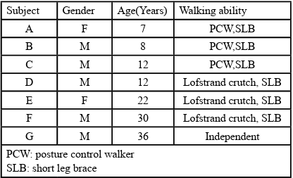
Table 1. Attributes of the subjects
The subjects were asked to walk back and forth on a 10 m flat surface using a Gait Corrector device. The pre- and after-walking protocols consisted of at least 5 minutes of rest before walking, followed by wearing a Gait Corrector device, light-load walking, and heavy-load walking based on the participant's rating of perceived exertion (RPE), followed by 10 minutes of rest, and then the end of the protocol. The exercise intensity was measured based on the heart rate while walking using a pulse oximeter (PMP-125, Pacific Medico Co. Ltd., Hong Kong), and none of the participants exceeded 20% of the load intensity.
Based on a report by Irie et al. [5], NIRS (OEG-16, Spectratech Inc.) electrodes were attached to the tibialis anterior and lateral gastrocnemius muscles on both sides without interfering with their movements (Fig.1-3). The blood oxygen dynamics of the local muscles were measured to confirm the degree of muscle fatigue after walking.
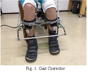
Fig. 1. Gait Corrector
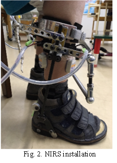
Fig. 2. NIRS installation
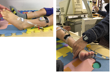
Fig. 3. Nerve conduction velocity measurement setting using evoked electromyography
Using NIRS, we measured the blood oxygen dynamics in local muscles from the resting state before walking to 10 minutes after walking. Based on our previous study [3,4], we analyzed the concentration changes (∆Hb-D) of oxygenated hemoglobin (∆Oxy-D) and reduced hemoglobin (∆Deoxy-D).
Suzuki et al. [5] used the maximum amplitude ratio of the F and M waves to clarify the degree of increased spastic muscle tone. The tone state of the lateral gastrocnemius muscle was measured three times before walking, immediately after heavy-load walking, and 10 minutes after walking, using an evoked potential testing device (Neuropack∑, MEB-5504, Nihon Kohden Co., Tokyo, Japan), with a recording electrode attached to the abductor digiti brevis behind the medial eminence. M- and F-waves were recorded by tibial nerve stimulation using a recording electrode on the abductor pollicis brevis muscle behind the medial phalanx and a reference electrode at the base of the big toe.
Sixteen measurements were performed at a maximum stimulation frequency of 0.5 Hz for the M-wave, and the average F/M ratio of 14 measurements, excluding the maximum and minimum values, was used as the F-wave parameter.
This study was conducted after explaining the study content to the participants in writing and obtaining their consent.
Results
Three participants who exhibited daily resistance to passive dorsiflexion of the ankle joint had a decrease in the mean F/M ratio immediately after walking but returned to the pre-walking state 10 minutes after the end of walking. Similar changes were observed after six months of walking (Fig. 4, Table 2).
Additionally, the blood oxygenation dynamics exhibited a greater disparity between oxyhemoglobin and deoxyhemoglobin concentrations after walking in all participants, suggesting local muscle fatigue, the degree of which showed no improvement even after 10 minutes of rest.
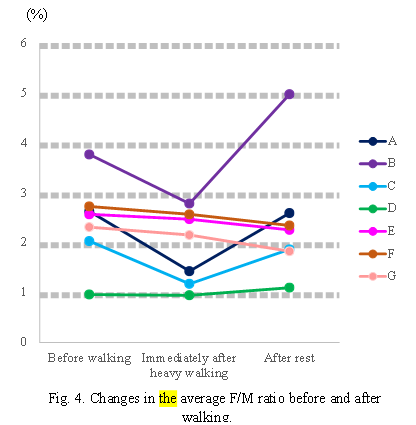
Fig. 4. Changes in the average F/M ratio before and after walking.
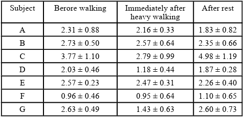
Table 2. Average F/M ratio before and after walking for each subject
Regarding the blood oxygen dynamics by NIRS, the difference between Oxy-D and Deoxy-D did not reveal a substantial change after light-load walking, and even when a temporary change was observed, it recovered to a steady state after approximately 5 minutes of rest.Conversely, after heavy-load walking, all of the participants exhibited disparity in the concentrations of oxyhemoglobin and deoxyhemoglobin, suggesting local muscle fatigue, and the degree of fatigue did not improve even after 10 minutes of rest. However, in participants with constantly hypertonic foot muscles, the difference between Oxy-D and Deoxy-D persisted even after 10 minutes of heavy-load walking, suggesting persistent local muscle fatigue (Fig. 5).
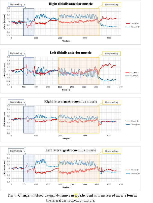
Fig. 5. Changes in blood oxygen dynamics in a participant with increased muscle tone in the lateral gastrocnemius muscle.
However, lateral gastrocnemius muscle fatigue has been rarely reported. In some participants whose joint range of motion was not limited by lower extremity muscle tension, the tibialis and lateral gastrocnemius muscles indicated non-fatigue conditions, as no difference between Oxy-D and Deoxy-D was observed immediately after heavy-load walking or at 10 minutes post-walking (Fig. 6).
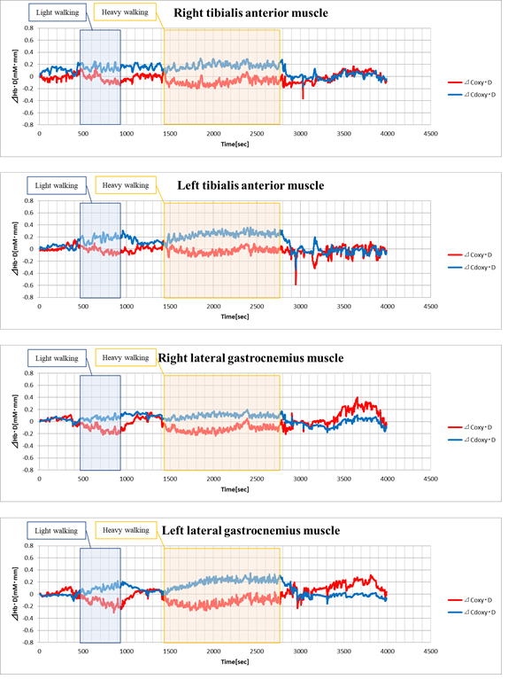
Fig. 6. Changes in blood oxygen dynamics in a participants with lower extremity muscle tone that does not limit the joint range of motion.
Discussion
Generally, children with cerebral palsy and muscle hypertonia in the lower extremities have increased tension in hip flexion, adduction, internal rotation, knee flexion, and pes equinovarus foot position during walking. This limits mobility and causes the lower extremities to cross over during stepping, making it difficult to achieve lower- extremity propulsion.
Although a Gait Corrector device allows step movement, a new mechanism called the link bar, which connects both legs to the front of the device, inhibits scissor foot (muscle tension of hip adduction and internal rotation), which causes the lower extremities to cross. This replaces the inhibited force with propulsion of the lower extremities and facilitates step widening.
Consequently, the support base area is secured, relieving tension in in the trunk and significantly improving gait. Furthermore, because the device is easy to use and can be used while sitting in a wheelchair or baby carriage, it is assumed that the tension in the legs gradually diminishes.
In this study, we conducted quantitative fatigue analysis after applying a Gait Corrector device in patients with cerebral palsy who were able to walk. Although the gait of the patients was objectively improved by walking practice as well as their own perception, it was confirmed that the degree of local muscle fatigue after walking, based on blood oxygenation dynamics and the quantitative changes in the electromyogram during walking, differed depending on the load intensity of walking and the degree of tension in the lower extremities. Surface electromyography (EMG) is the most commonly used noninvasive method for assessing local muscle fatigue. However, the magnitude of surface electromyography is relative, and muscle fatigue can deviate significantly from the actual muscle fatigue depending on the measurement conditions and measurement site.
NIRS is an optical measurement method that uses near-infrared rays to measure blood oxygenation noninvasively. Typically, NIRS-based products are used to visualize brain activity within the skull. There have been several reports on the application of NIRS to muscular regions but not to the brain [6-10].
Hamaoka et al. [6,7] introduced a noninvasive measurement of muscle metabolism by combining magnetic resonance spectroscopy, which can calculate the concentration of substances directly involved in the biochemical reactions of cells, and NIRS, which can assess the oxygenation state of intravascular hemoglobin. Additionally, DeLorey et al. [8] and Grassi et al. [9] have suggested that changes in deoxygenated hemoglobin concentration measured using NIRS reflect a balance between oxygen transport and utilization at the gas exchange vascular level. Building on their work, Ferreira et al. [10-12] have published several studies showing that the dynamics of the deoxygenated hemoglobin concentration reflect the dynamics of the arteriovenous oxygen range. Saitoh et al. [13] also found that NIRS measurements were not affected by changes in the blood flow velocity. They also reported that the dynamics of the deoxygenated hemoglobin concentration reflected the dynamics of the arteriovenous oxygen range. Muramatsu et al. [14] conducted various experiments to investigate the relationship between local fatigue and blood oxygen dynamics. They also reported that the difference between oxygenated and deoxygenated hemoglobin levels increases with fatigue.
Generally, even children with cerebral palsy who acquire gait at an early age lose the ability to walk over time because of continuous walking, which causes abnormal muscle tone in the lower extremities, affecting the bones and joints. In adults, it is extremely difficult to maintain walking because of the effects of proportional changes.
The Gait Corrector device is expected to meet the needs of children with cerebral palsy who wish to walk without upper extremity support, including a cane, or specifically, "walk" in their natural bipedal state, and help them achieve an efficient gait that cannot be achieved with conventional walkers or orthoses. If children with cerebral palsy can relearn their sense of balance while walking through the application of a gait corrector and maintain their ability to walk in the long term, it will enhance their quality of life in terms of daily activities.
In conclusion, a quantitative fatigue analysis was performed in patients with cerebral palsy who were able to walk using a Gait Corrector device. Compared with conventional short-leg orthoses, the Gait Corrector is a device that suppresses muscle tone during hip adduction and internal rotation, and it is capable of replacing the suppressed force with a propulsive force in the lower extremity. The gait of the patients was objectively improved by walking practice. However, the degree of local muscle fatigue after walking in terms of blood oxygen dynamics differed depending on the load intensity of walking and the degree of tension in the lower extremities.
In addition to being small, lightweight, and easy to operate, NIRS can noninvasively observe fatigue in local muscles and objectively capture the effects of clinical training. In addition, NIRS can measure post-exercise conditions that cannot be captured by electromyography. It is easy to evaluate fatigue recovery after training using blood oxygen dynamics, which is expected to be useful in the rehabilitation field.
Competing Interests:
The authors declare that they have no competing interests for this study.
Acknowledgments:
The authors are grateful to the participants and co-authors for their assistance with the data acquisition.
References
Kazutaka, I., Ryo, T., Ryuji, Y., & Masaharu, M., (2006). Function of Hart Walker and its effect for independent mobility. Bulletin of the Japanese Society of Prosthetics and Orthotics, 22(2), 90–94.
Hiroshi, K., Takeo, K., So Nakayama, & Kazutaka, I., (2008). Clinical trial of an active walker that enables walking even with total paralysis. Nervous System in Children 33, 101–106.
Masaharu, M., Yoshiki, M., Kobayashi, H., & Kazutaka, I., (2013). Trial of fatigue evaluation in cerebral palsy by near infrared spectroscopy. Journal of the Japanese Physical Therapy Association, 25, 8 (Suppl. 1).
Masaharu, M., Masafumi, K., Yu, I., Yoshihiro, K., & Kazutaka, I., (2016). Evaluation of the local muscle in cerebral palsied children using gait corrector, 19th International Meeting of Physical Therapy Science, Silla University, Busan, South Korea, July 16(suppl).
Hirofumi, W., (2002). Walking and gait: Journal of Kansai Physical Therapy, 2, 41–44.
Takafumi, H., & Takayuki, S., (2003). Establishment of noninvasive measures of skeletal muscle circulation and metabolism: Application to Angiology. The Journal of Japanese College of Angiology, 43(6), 239–244.
Toshihito, F., & Takafumi, H., (1998). Non-invasive metabolic measurement. Japanese Journal of Physical Fitness and Sports Medicine, 47(3), 367–372.
DeLorey, D. S., Kowalchuk, J. M., Paterson D. H. (2003). Relationship between pulmonary O2 uptake kinetics and muscle deoxygenation during moderate-intensity exercise. Journal of Applied Physiology, 98, 113–120.View
Grassi, B., Pogliaghi, S., Rampichini, S., Quaresima, V., Ferrari, M., Marconi, C., & Cerretelli, P., (2003). Muscle oxygenation and pulmonary gas exchange kinetics during cycling exercise on transitions in humans. Journal of Applied Physiology, 95, 149–158.View
Ferreira, L. F., Townsend, D. K., Lutjemeier, B. J., Barstow, T. J. (2005). Muscle capillary blood flow kinetics estimated from pulmonary O2 uptake and near-infrared spectroscopy. Journal of Applied Physiology, 98, 1820–1828.View
Ferreira, L. F., Poole, D. C., Barstow, T. J., (2005). Muscle blood flow-O2 uptake interaction and their relation to on exercise dynamics of O2 exchange. Respiratory Physiology & Neurobiology, 147, 91–103.View
Ferreira, L. F., Harper, A. J., Townsend, D. K., Lutjemeier, B. J., Barstow, T. J., (2005). Kinetics of estimated human muscle capillary blood flow during recovery from exercise. Experimental Physiology, 90, 715–726.View
Tadashi, S., Tadanori, F., Hirotaka, Y., Tatsuhisa, T., Kyuichi, N., (2011). Measurement of hemoglobin concentration by Near infrared spectroscopy is independent of changes in blood flow velocity. Transactions of Japanese Society for Medical and Biological Engineering, 49(1), 185–190.View
Yoshiki, M., & Hiroshi, K., (2011). Assessment of muscle fatigue by NIRS. Transactions of the JSME (in Japanese), 80(814), 559–567.View