
- About the Journal
- Editorial Board
- Review Process
- Author Guidelines
- Article Processing Charges
- Special Issues
- Current Issue
- Past Issue
 Current Research in Materials Chemistry
Current Research in Materials Chemistry Volume 4 (2022), Article ID: CRMC-113
https://doi.org/10.33790/crmc1100113Research Article
Dissolution of Kidney Stones; Nano and Micro Morphologies Developed During Remelting
Abhishek Singh, Krishna S. Machuga, Himagowri Prasad, Kamdeo Mandal*, Lisa Kelly, Bradley Arnold, Fow-Sen Choa, Brian Cullum, Glenn Austin** and N. B. Singh
Department of Chemistry and Biochemistry, University of Maryland Baltimore County, 1000 Hilltop Circle, Baltimore, MD 21240, United States
*Corresponding Author Details: Kamdeo Mandal, Professor, Department of Chemistry, Indian Institute of Technology, Banaras Hindu University, Varanasi (UP) India.
**Co-Author: Glenn Austin, Department of Chemistry and Biochemistry, University of Maryland Baltimore County, 1000 Hilltop Circle, Baltimore, MD 21240, United States. E-mail: kdmandal.apc@itbhu.ac.in
Received date: 10th January, 2022
Accepted date: 17th January, 2022
Published date: 19th January, 2022
Citation: Singh, A., Machuga, K. S., Prasad, H., Mandal, K., Kelly, L., Arnold, B., Choa, F. S., Cullum, B., Austin, G., & Singh, N.B. (2022). Dissolution of Kidney Stones; Nano and Micro Morphologies Developed During Remelting. Cur Res Mate Chem 4(1): 113.
Copyright: ©2022, This is an open-access article distributed under the terms of the Creative Commons Attribution License 4.0, which permits unrestricted use, distribution, and reproduction in any medium, provided the original author and source are credited.Creative Commons Attribution License, which permits unrestricted use, distribution, and reproduction in any medium, provided the original author and source are credited.
Abstract
We have synthesized calcium oxide and calcium oxalate based kidney stones and investigated remelting to understand the process of dissolution. The dissolution morphology and remelting process in water was observed for the pure and intensely impurity doped oxalate-urate stones grown at lower pH to study effect of acidity, and pH near actual pH of the stomach. Direct observation showed different melting morphologies. We observed needles, plates, dendrites, and lamella depending on the pH level and impurities. Remelting studies indicated that during breaking of large polycrystalline bunched stones grown at lower pH break into faceted small crystals which dissolve into the solvent depending on the acidity pH and impurities. High purity stones grown near stomach pH (6.4-7.5) tends to grow in needles morphology which dissolve slowly and fragment into smaller needles. A comparison was made with real kidney stones observed at Herring laboratory and similarity was observed with impurity doped stones. There was significant difference in decomposition of pure and impurity doped stones. Thermal analysis (DTA) showed that sugar doped oxides decompose continuously.
Keywords: Crystal, Growth, pH, Needles, Prism, Kidney, Stones, Crystallization, Coarsening
Introduction
There is a continuous effort to decrease cost of industrially important materials specifically bulk, thin film and nano structured materials. This puts more thrust to explore multifunctionality in materials. Recently some progress has been made in sulfides, selenides and oxides which are very useful in optical, electronic and energy storage industries. Some progress has been made in chalcogenides and chalcopyrites [1,2] which have shown applications in laser developments and hyperspectral imagers and multinary oxides [3-5] for dielectric energy storage and insulation layers. With evolution of nano scale structures, some of the materials properties have evolved which are dependent on morphologies. These studies demonstrate some highlights on transition of micro and nano particles into nano cubes, paraboloids, nanowires, and other shapes. This also helps in explaining kinetics and mechanism of different morphologies observed for kidney stones. Figure 1 shows transition of nanoparticles to form nanowires. The analysis of real human kidney stones by Glenn Austin of Louis C. Herring Corporation have shown [6] that hundreds and thousands of analysis of kidney stones show variety of shapes sizes and colors.
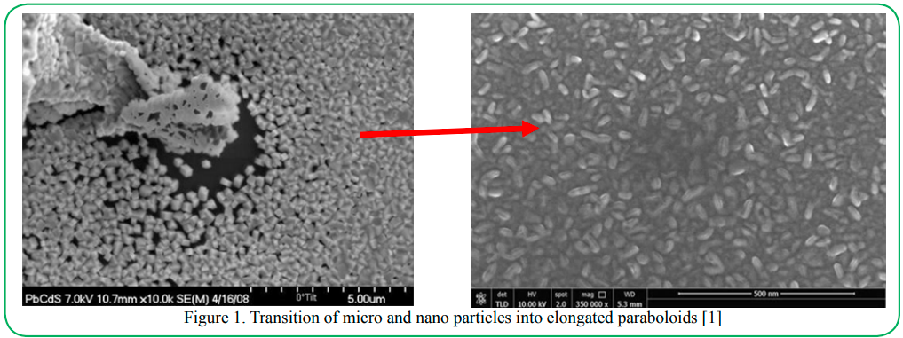
Figure 1. Transition of micro and nano particles into elongated paraboloids [1]
Although growth of kidney stones in stomach has doubled in the world’s population in past several decades, there is not a single reason for its formation [6-8]. But it is suggested that the increasing exposure to antibiotics and antacids are few causes. The typical morphologies reported by the Herring Lab are very interesting. We had shown [9,10] that the effects of pH, sugar and MgO has significant effect on the formation and morphology of stones. In this paper we performed experiments to remelt the synthesized oxalate-urate based kidney stones to understand the mechanism of formations from nano and micro particles.
2. Experimental Methods
2.1 Materials Synthesis: The typical kidney stones contain approximately calcium oxalate (carbonate) 75%, calcium phosphate 5%, mixed oxides 5%, uric acid 1-10%, cystine (C6H12N2O4S2) 0.1 to 1.5%, Miscellaneous metal salts and oxides >1.0% and sugar 0.0.01%. To synthesize the kidney stone in the laboratory, we used calcium carbonate (CaCO3), calcium oxalate listed for 99.9%, urea (NH2CoNH2) 99.99% and glucose 98% (sugar). All chemicals were purchased from Sigma Aldrich and no further purification was performed. Sodium from the sodium hydroxide was used as the nutrient for sodium during synthesis. Typical mixture contained ratio in weight percentage of CaCO3, Urea, and sodium salt in 70:20:10 ratio. The amount of sugar was small (10mg). These materials were mixed and dissolved in dilute acetic acid. The pH was controlled by adding solution of sodium hydroxide (NaOH) and acetic acid. Few drops ( 2-4 cc) of nitric acid was also required to completely dissolve the calcium oxides. In several experiments we added 1% TiO2also.
2.2 Growth of stones: Stones in multi crystalline states were grown by solution growth method in the water-acetic acid solvent. The solvent contained a 3:1 ratio of water to acetic acid. Typical runs were in 150 to 200 cc solvent. NaOH solution and acetic acid were used in the solvent to control the pH. The solvent containing mixture was heated to 80C to completely dissolve the mixture and to achieve transparent solution. The temperature was lowered to 45o C and kept for few hours to achieve homogeneous solution.
3. Result and Discussion
3.1 Growth of undoped urate stones: Effect of pH and impurity on the growth morphology have significant effect on the morphology of kidney stones [9-11]. The size and crystallinity also depended on the cooling rate and pH of the solution. Nano, micro and cm size multi crystalline solids were observed by quenching or slow cooling. Fast quenching in ice of the solution produced small size bunched materials (Figure 2a). Self - cooling from 45oC, to room temperature produced uncontrolled nucleation (Figure 2b). In this process, white precipitates between needles were also observed similar to previous observations for other materials [9-13] for oxides and vanillin. These needles attached to polycrystalline precipitates of very small sizes (Figure 3 and 4). Due to the fast cooling and fast growth rate, crystals harvested were not completely transparent and had striations.
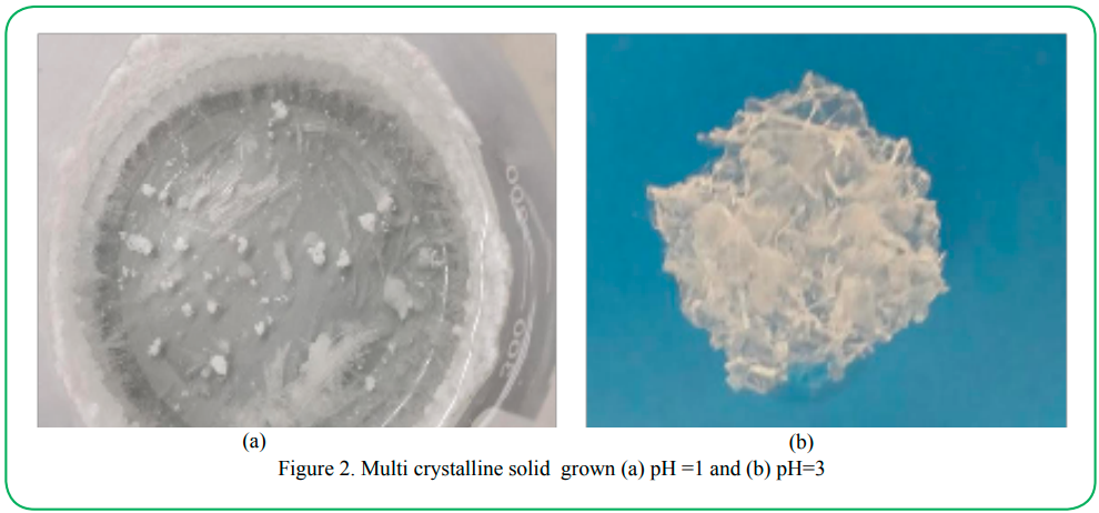
Figure 2. Multi crystalline solid grown (a) pH =1 and (b) pH=3
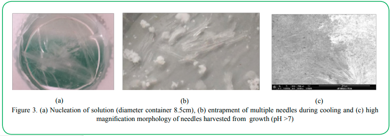
Figure 3(a). Nucleation of solution (diameter container 8.5cm), (b) entrapment of multiple needles during cooling and (c) high magnification morphology of needles harvested from growth (pH >7)
3.2 Remelting phenomena: We performed remelting experiments similar to that reported earlier [12] by raising the temperature to understand the dissolution of kidney stones. This study was focused to evaluate the characteristics of dissolution of high purity stones at low and high pH. Figure 3 shows the re-melting characteristics of stones grown at low pH values. This clearly indicates that during breaking of kidney stones the joining material breaks first leaving the large, faceted crystals which are undissolved in the beginning. With increasing time or lowering pH (closer to 1) these structires break and then dissolve into very small crystallites. Figure 4 shows that there is no sign of layered growth or growth steps in large needles. The crystal growth of calcium urate by Swift et al [13] indicated needle morphology but much higher aspect ratios. Melting showed that small polycrystalline translucent material which grows between needles, melts first and large needles do not break at early stage of melting. As reported in reference [9-12] for kidney stone material and vanillin, the needles observed were very small and fat. Compared to that obtained by Swift et al [13]. Based on reported morphology of kidney stones by Herring laboratory, it appears that the needles with very high aspect ratio are unrealistic for human kidney stones. Experiments of Swift et al. was performed with the high purity calcium urate and that may be the reason for very large needles. As has been reported in the real situation, it is expected that real kidney stones are contaminated due to varieties of impurities such as sodium, sugar, metallic and nonmetallic impurities. Since impurities affect the morphology significantly, needles can easily change (Figure 5) into plates or dendrites. Because of this reason we have added sugar and magnesium doped stones and compared with real kidney stones in following section.
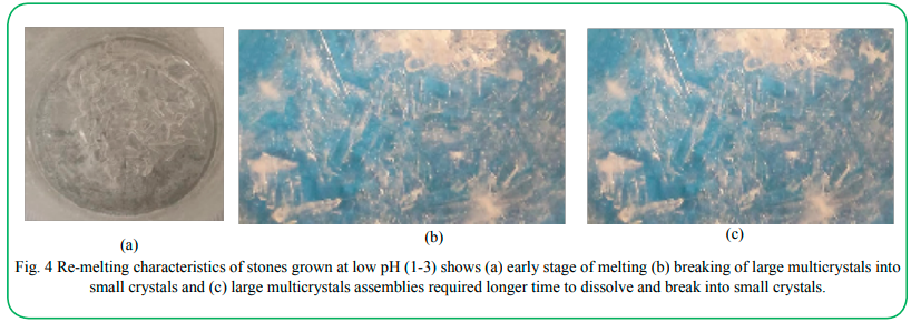
Figure 4. Re-melting characteristics of stones grown at low pH (1-3) shows (a) early stage of melting (b) breaking of large multicrystals into small crystals and (c) large multicrystals assemblies required longer time to dissolve and break into small crystals.
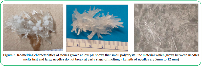
Figure 5. Re-melting characteristics of stones grown at low pH shows that small polycrystalline material which grows between needles melts first and large needles do not break at early stage of melting. (Length of needles are 5mm to 12 mm)
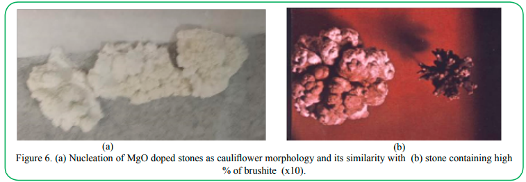
Figure 6.(a) Nucleation of MgO doped stones as cauliflower morphology and its similarity with (b) stone containing high % of brushite (x10).
2.5 Effect of impurities: Both organic and inorganic impurities affect the kinetics and morphology of growth and crystals significantly. In most of the cases impurities decrease the growth rate and modify the nucleation as well as nano and micro morphology. Since variety of impurities are filtered by the real kidney including magnesium and sugar, we doped the original solution with 5 wt% MgO and in another experiment MgO and sugar. A typical morphology of Mg ion doped crystal is shown in Figure 6. It showed a cauliflower morphology similar to that obtained for struvite (ammonium magnesium phosphate). The mechanism of the modification is very complex since it affects the growth interface, segregation and hence magnesium changed the nucleation and growth morphology. It is appears that the growth starts with nucleation forming spherulites, but further study is required. Figure 7 shows that morphology consists of many small nano and microcrystals arranged like a cauliflower morphology. Also, these structures are very hard compared to un doped material. Effect of sugar was studied by adding sugar (C6H12O6) by adding sugar (<1%) in the solution. This resulted in brownish stones with huge number of nano and microcrystals jointed together. This study was focused to evaluate effect at room temperature for a longer time (more than 100 hours) effect. We placed the solution for a time period of 150 hours for the nucleation.
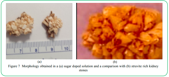
Figure 7: Morphology obtained in a (a) sugar doped solution and a comparison with (b) struvite rich kidney stones
Figure 7 shows the effect of sugar on the colorization and formation of surface morphology. It consisted of small crystallites oriented randomly. The detailed decomposition studies using differential thermal analysis is discussed in reference 10. The sugar containing sample started decomposing at low temperature and showed some steps in the decomposition curves. This step decrease indicates that during decomposition sugar (C6H12O6) containing stones may cause formation of other side products at embodied in the stones. Thermal analysis (DTA) showed that sugar doped oxides decompose continuously, and this study is continuing.
4. Conclusion
The remelting process in water was studied for the pure and intensely impurity doped oxalate-urate stones grown at lower and higher pH. Direct observation showed different melting morphologies since different pH and impurities had produced needles, plates, dendrites, and lamella depending on pH and impurities. Remelting studies indicate that stones grown at lower pH break into faceted crystals which dissolve into the solvent depending on the acidity pH and impurities. Stones grown near stomach pH (6.4-7.5) in needles morphology which break into smaller needles. A comparison with morphology of real kidney stones observed at Herring laboratory showed similarity in morphologies.
Acknowledgement
The authors would like to acknowledge and thank Herrings Laboratory, Orlando for his permission to use pictures of real kidney stones. The authors are also grateful to Northrop Grumman for providing the chemicals for this project.
Competing interests:
The authors report no conflict of interest.
References
Narsingh Bahadur Singh, Christopher Cooper, Pietro Strobbia, Narasimha Prasad, Ching Hua Su, Bradley Arnold, Fow-Sen Choa, (2017). "Nanomorphology and performance of pure and doped lead selenide for infrared detector," Opt. Eng. 56(7), 077106View
N. B. Singh, Ching-Hua Su, Brad Arnold and Fow-Sen Choa. Stacey Sova and Christopher Cooper, (2017). “Multifunctional 2D Materials; Gallium Selenide, Materials Today 4 ,5471-5477.View
Laxman Singh, U. S. Rai, K. D. Mandal, and N. B. Singh, (2014). “Progress in the growth of CaCu3Ti4O12and related functional dielectric perovskites”, Progress in Crystal Growth and Characterization 60,15-62.View
N. B. Singh, S.R. Coriell, R. H. Hopkins, Ching Hua Su, B. Arnold, Fow-Sen Choa and Brian Cullum, (2014). “Growth mechanism of nanowires: Binary and ternary chalcogenides”. Nanoscience and Technology, 1, 2.1-8View
N. B. Singh, A. Berghmans, D. J. Knuteson, John Talvacchio, D. Kahler, M. House, B. Schrieb, B. Wagner and M. King, (2012). “Evolution of microstructure due to additives and processing”, Materials Science and Technology Conference 2011,Edited by Priya Shashank and K. M. Nair, Advances in applications in electronics II ,65-75. View
Glenn Austin, Integrated approach to kidney stone analysis”, Louis C. Herring Corporation (Private Communication) July 2018.
Robert H. Shmerling, MD, (2016). “Kidney stones are on the rise”, Harvard Health Publishing, Harvard Medical School, Feb 12.View
Silvia De Rosa Massimo Antonelli Claudio Ronco, (2017). “Hypothermia and kidney: a focus on ischaemia–reperfusion injury”, Nephrology Dialysis Transplantation, 32, 2, Pages 241–247.View
Neelesh Agarwal, Stacey Sova, N. B. Singh, Brad Arnold, Fow-Sen Choa, Brian Cullum and Ching-Hua Su, (2016). “Effect of pH on the morphology of kidney stones”, Proc. SPIE 9863, Smart Biomedical and Physiological Sensor Technology XIII, Brian M. Cullum; Douglas Kiehl; Eric S. McLamore, Editor(s) 986303, 13 May.View
N. B. Singh, Ching Hua Su, Fow-Sen Choa, Brian Cullum, Brad Arnold, Lisa Kelly, Neelesh Agarwal, Stacey Sova and Christopher Cooper, (2019). “Effect of sugar and pH on the morphology, bulk transparency and thermal characteristics of kidney stones”, Journal of Scientific Research 9(1) , 11-18.
O. P. Singh, Namwar Singh, Y.P. Singh, and N. B. Singh, (2001). “Growth of vanillin crystals for second harmonic generation applications in near-IR wavelength region J. Crystal Growth 225(2) 470-73.View
N. B. Singh, T. Henningsen, E. P. A. Metz, R. Hamacher, E. Cumberledge, R. H. Hopkins, and R. Mazelsky, (1991). “Solution growth of vanillin crystals”, Materials Letter, 12(4) 270-275.View
J. B. Presores, K. E. Cromer, Christina Capacci-Daniel and Jennifer A. Swift, (2013). Calcium Urate hexahydrate, Cryst. Growth Des. 13, 5162−5164.View